Copyright
©The Author(s) 2021.
World J Clin Cases. Aug 16, 2021; 9(23): 6781-6788
Published online Aug 16, 2021. doi: 10.12998/wjcc.v9.i23.6781
Published online Aug 16, 2021. doi: 10.12998/wjcc.v9.i23.6781
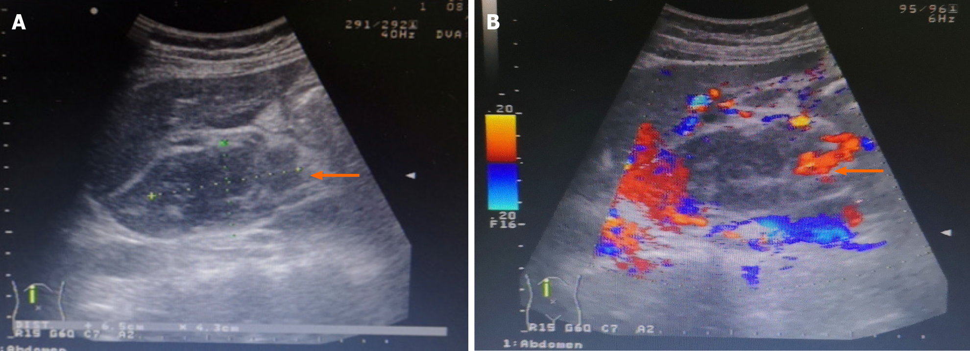
Figure 1 Ultrasound examination of the liver and gallbladder.
A: A hypoechoic mass is seen in the caudate lobe of the liver, being about 6.5 cm × 4.3 cm in size, with blurred boundary and irregular shape; B: No obvious blood flow signal is seen in this mass, which had prompted consideration of the possibility of a malignant tumor.
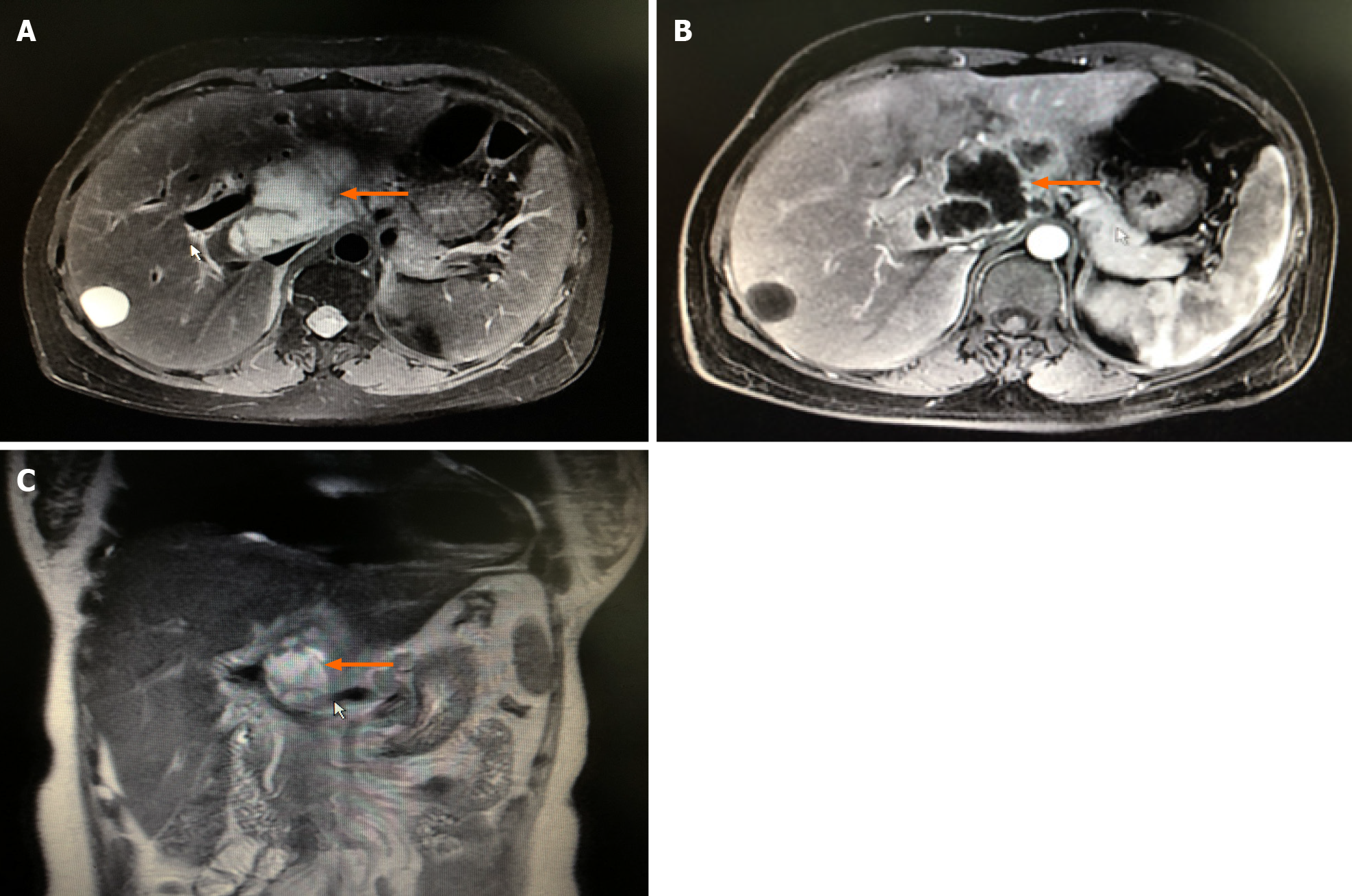
Figure 2 Enhanced magnetic resonance imaging of the liver.
A and C: A space-occupying lesion (orange arrows) is seen axially (A) and coronally (C) in the caudal lobe of the liver, with unclear borders and irregular shape, about 7.6 cm × 4.4 cm × 5.0 cm in size, slightly low signal on T1WI and slightly high signal on T2WI, and inhomogeneous signal; B: The edge of the tumor is enhanced in the arterial phase, with multiple small and disorganized vascular shadows present within the lesion. Considering the possibility of cystadenocarcinoma, it was considered in differential diagnosis of liver abscess.
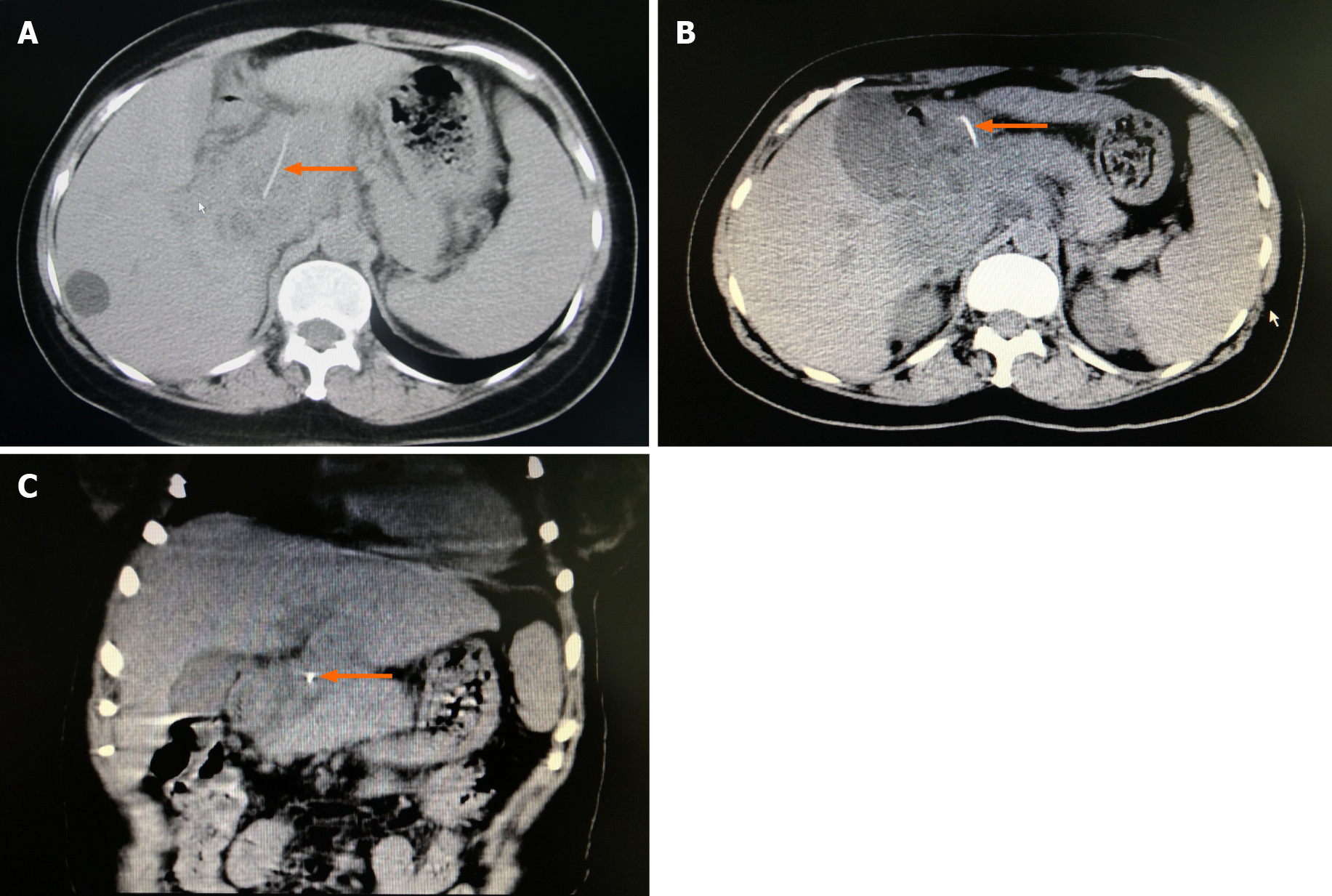
Figure 3 Computed tomography scan of the liver, gallbladder, and spleen.
A and B: An irregular soft tissue density shadow with poorly defined borders (orange arrow) is seen above the hilum, caudate lobe, and pancreatic head. A more hypodense focus is seen inside (orange arrow), measuring about 7.8 cm × 6.0 cm × 5.0 cm. A dense shadow of about 3.7 cm in length is seen inside the lesion, the anterior end of which is located in the gastric cavity; C: A foreign body (fishbone) is seen in the upper abdomen (orange arrow) and was considered to have penetrated the gastric wall to the hepatic hilum, with an abscess having formed above the caudal lobe and pancreatic head.
Figure 4 Gastroscopic view.
A: The mucosa of the anterior wall of the duodenal bulb is shown to be rough and uneven, with small bubbles throughout; B: Milky white pus is seen exudating from the anterior wall of the duodenal bulb, and the foreign body was considered to have punctured the anterior wall of the duodenal bulb.
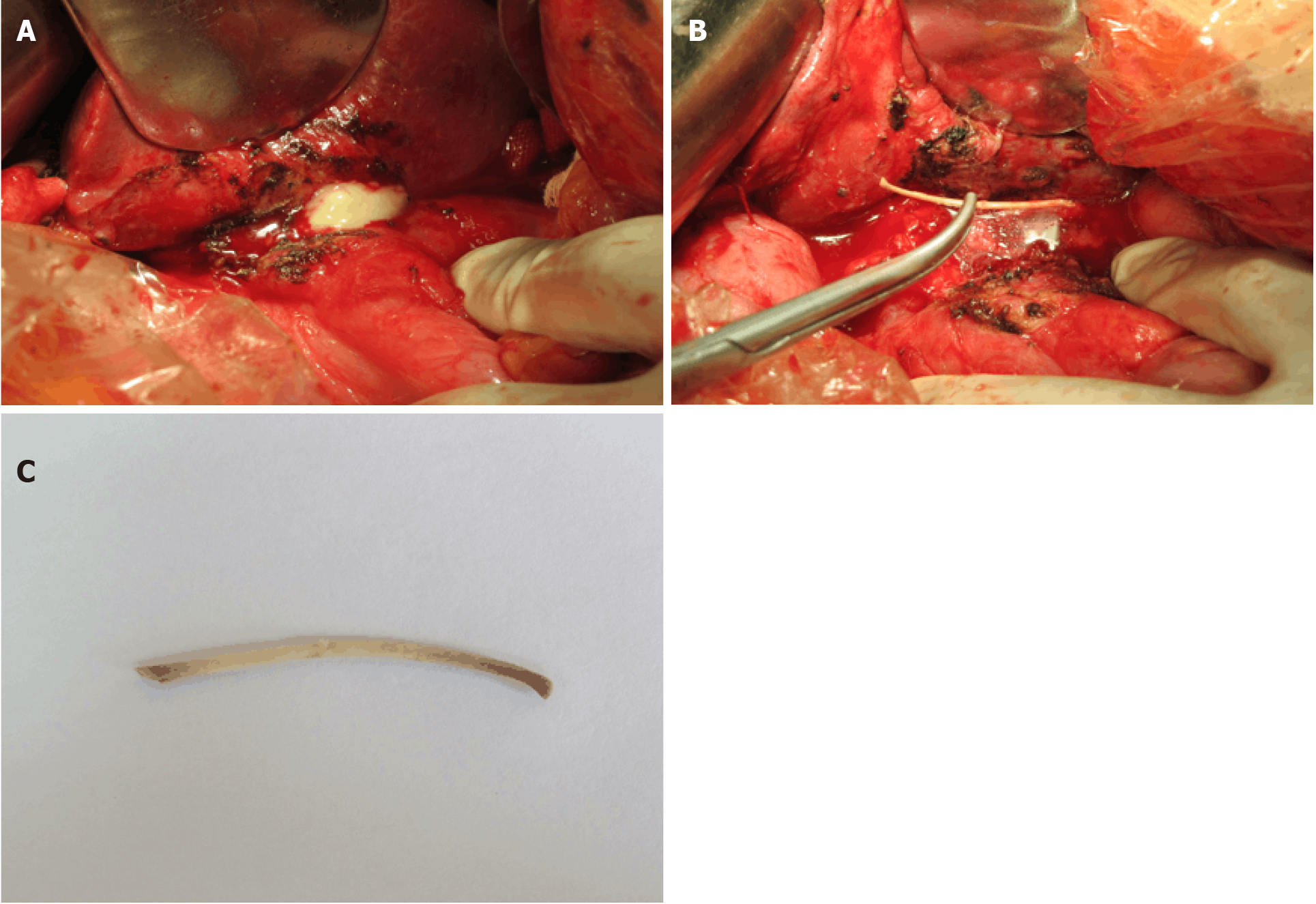
Figure 5 Intraoperative gross findings.
A: A large multi-lumen thick-walled abscess measuring approximately 9.0 cm × 8.0 cm × 8.0 cm is seen anterior to the caudate lobe of the liver and superior to the head of the pancreas, with no bony foreign body seen within it; B: Ultrasonographic imaging showing an additional abscess cavity deep within the large abscess, in which a foreign body, seen here grossly, was found deep behind the portal vein, measuring about 5.0 cm × 4.0 cm × 4.0 cm; C: Upon removal, the bony foreign body was identified as a fishbone about 4 cm in length and 0.3 cm in diameter.
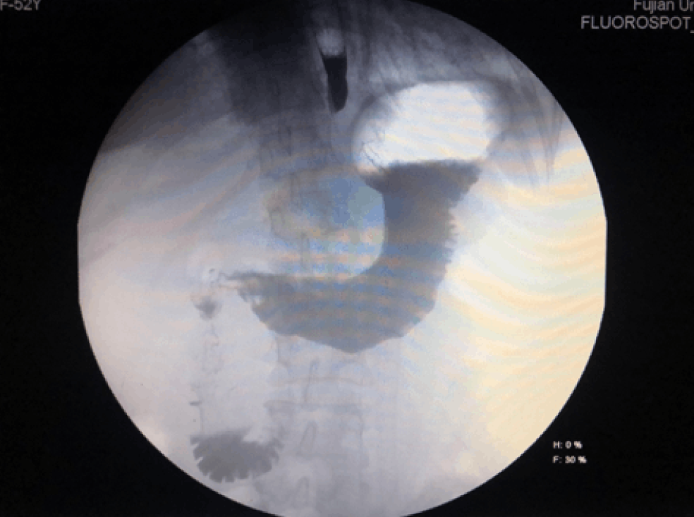
Figure 6 Digital gastroenterography view at 2 wk postoperative follow-up.
No contrast extravasation was observed in the gastroduodenum.
- Citation: Pan W, Lin LJ, Meng ZW, Cai XR, Chen YL. Hepatic abscess caused by esophageal foreign body misdiagnosed as cystadenocarcinoma by magnetic resonance imaging: A case report. World J Clin Cases 2021; 9(23): 6781-6788
- URL: https://www.wjgnet.com/2307-8960/full/v9/i23/6781.htm
- DOI: https://dx.doi.org/10.12998/wjcc.v9.i23.6781


