Copyright
©The Author(s) 2021.
World J Clin Cases. Aug 16, 2021; 9(23): 6775-6780
Published online Aug 16, 2021. doi: 10.12998/wjcc.v9.i23.6775
Published online Aug 16, 2021. doi: 10.12998/wjcc.v9.i23.6775
Figure 1 Positron-emission tomography/computed tomography image revealed a left seminal mass on baseline examination (A) and on contrast (B).
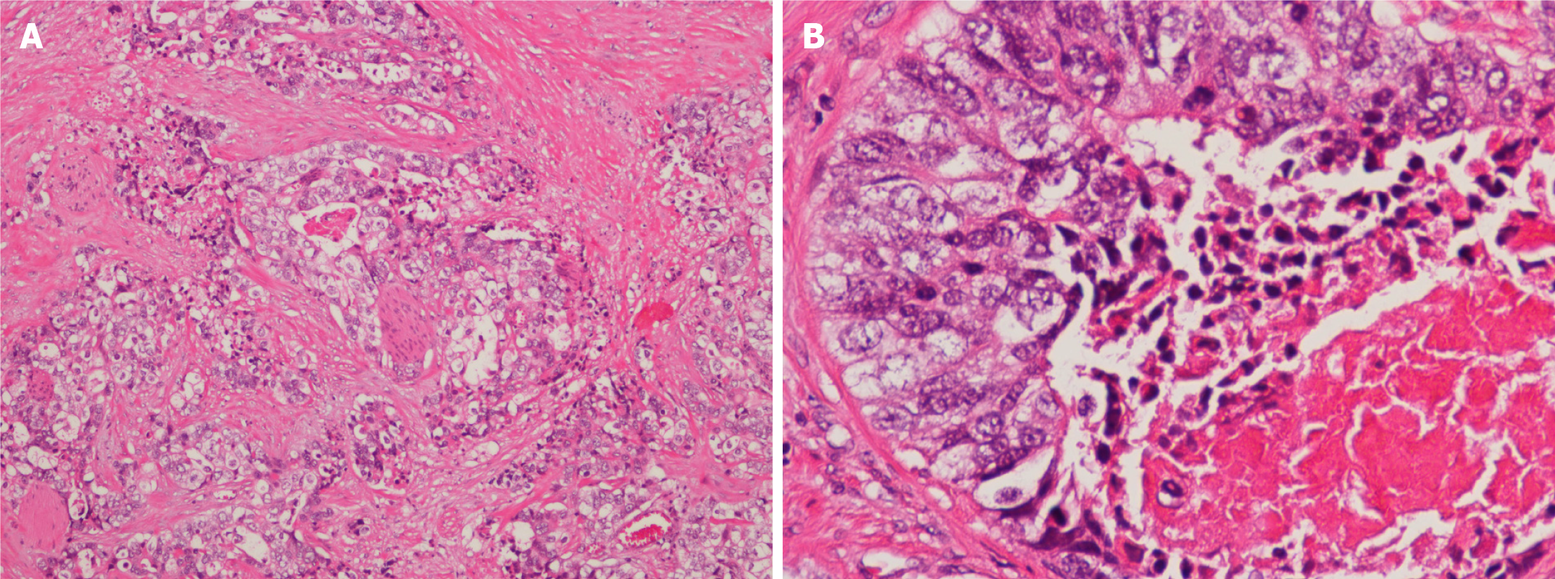
Figure 2 Microscopic view of the tumor in the seminal vesicle: The tumor cells were arranged in trabecular and solid nests, which was compatible with the original small bowel adenocarcinoma.
A: HE staining, 100 × magnification; B: HE staining, 400 × magnification.
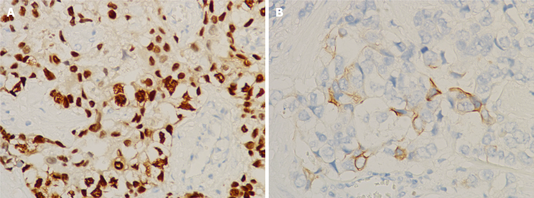
Figure 3 The tumor cells from the seminal vesicle were positive for CDX-2(++++) (A) and CK20(+) (B).
A: CDX2 staining, 400 × magnification; B: CK20 staining, 400 × magnification.
- Citation: Cheng XB, Lu ZQ, Lam W, Yiu MK, Li JS. Solitary seminal vesicle metastasis from ileal adenocarcinoma presenting with hematospermia: A case report. World J Clin Cases 2021; 9(23): 6775-6780
- URL: https://www.wjgnet.com/2307-8960/full/v9/i23/6775.htm
- DOI: https://dx.doi.org/10.12998/wjcc.v9.i23.6775


