Copyright
©The Author(s) 2021.
World J Clin Cases. Aug 16, 2021; 9(23): 6768-6774
Published online Aug 16, 2021. doi: 10.12998/wjcc.v9.i23.6768
Published online Aug 16, 2021. doi: 10.12998/wjcc.v9.i23.6768
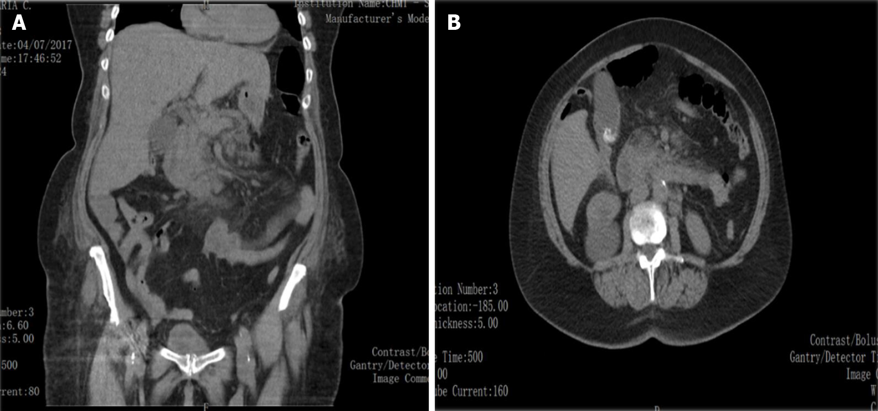
Figure 1 Computed tomography scan performed in the Emergency Department showing gallstones and changes in the pancreas and pancreatic fatty infiltration.
A and B: Coronal plane (A) of computed tomography scan and axial plane (B) showing minimum peritoneal leaking, gallstones with no biliary tract dilation and heterogeneous contour of the pancreas, with focal pancreatic fatty infiltration.
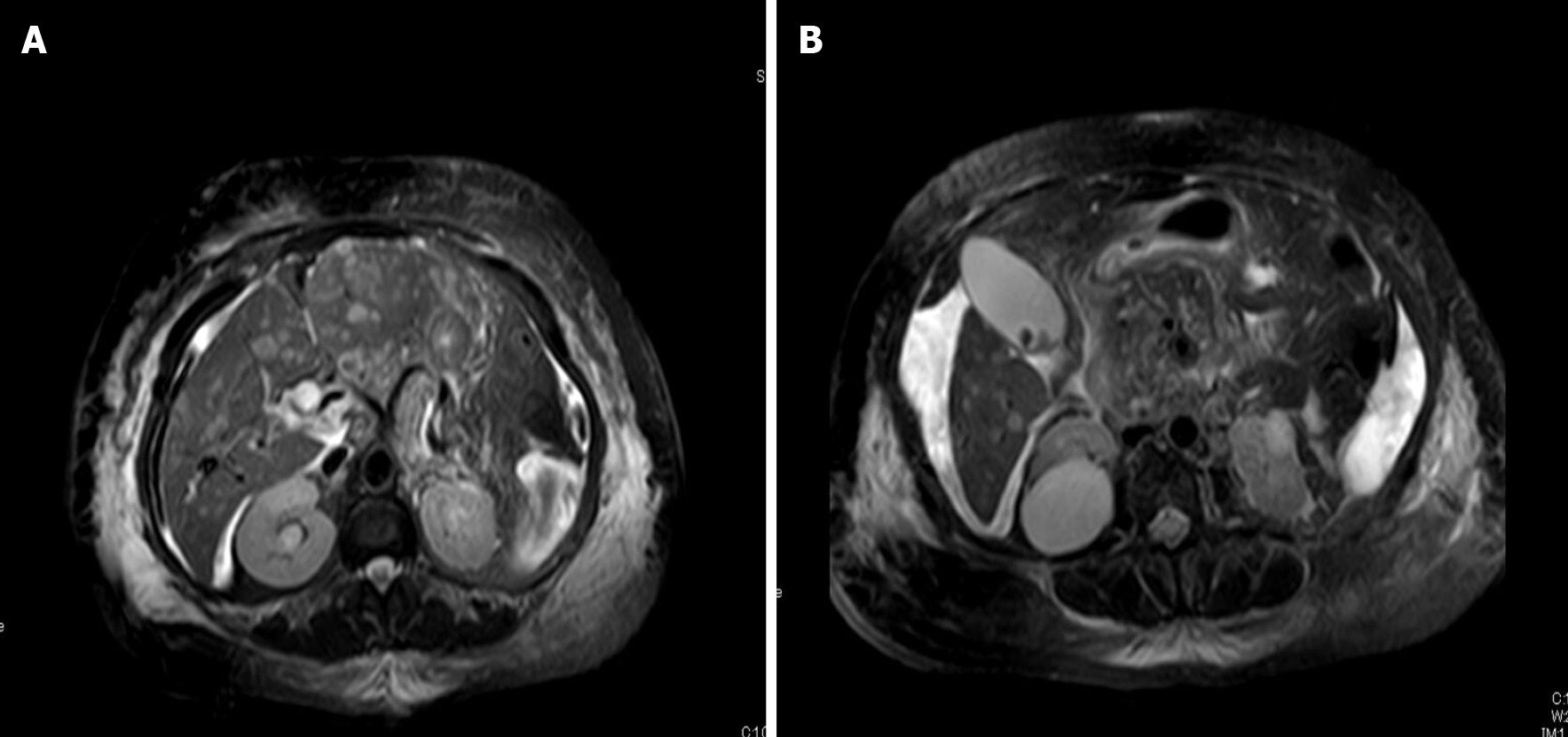
Figure 2 Magnetic resonance cholangiography showing globosity of the pancreatic head and changes in liver parenchyma.
A and B: Both Axial images showing multifocal liver lesions (A) and pancreatic heterogeinity (B).
- Citation: Bezerra S, França NJ, Mineiro F, Capela G, Duarte C, Mendes AR. Pylephlebitis — a rare complication of a fish bone migration mimicking metastatic pancreatic cancer: A case report. World J Clin Cases 2021; 9(23): 6768-6774
- URL: https://www.wjgnet.com/2307-8960/full/v9/i23/6768.htm
- DOI: https://dx.doi.org/10.12998/wjcc.v9.i23.6768


