Copyright
©The Author(s) 2021.
World J Clin Cases. Aug 6, 2021; 9(22): 6557-6565
Published online Aug 6, 2021. doi: 10.12998/wjcc.v9.i22.6557
Published online Aug 6, 2021. doi: 10.12998/wjcc.v9.i22.6557
Figure 1 The tip positions of the umbilical arterial catheter/umbilical venous catheter were in the 6th-7th thoracic vertebra.
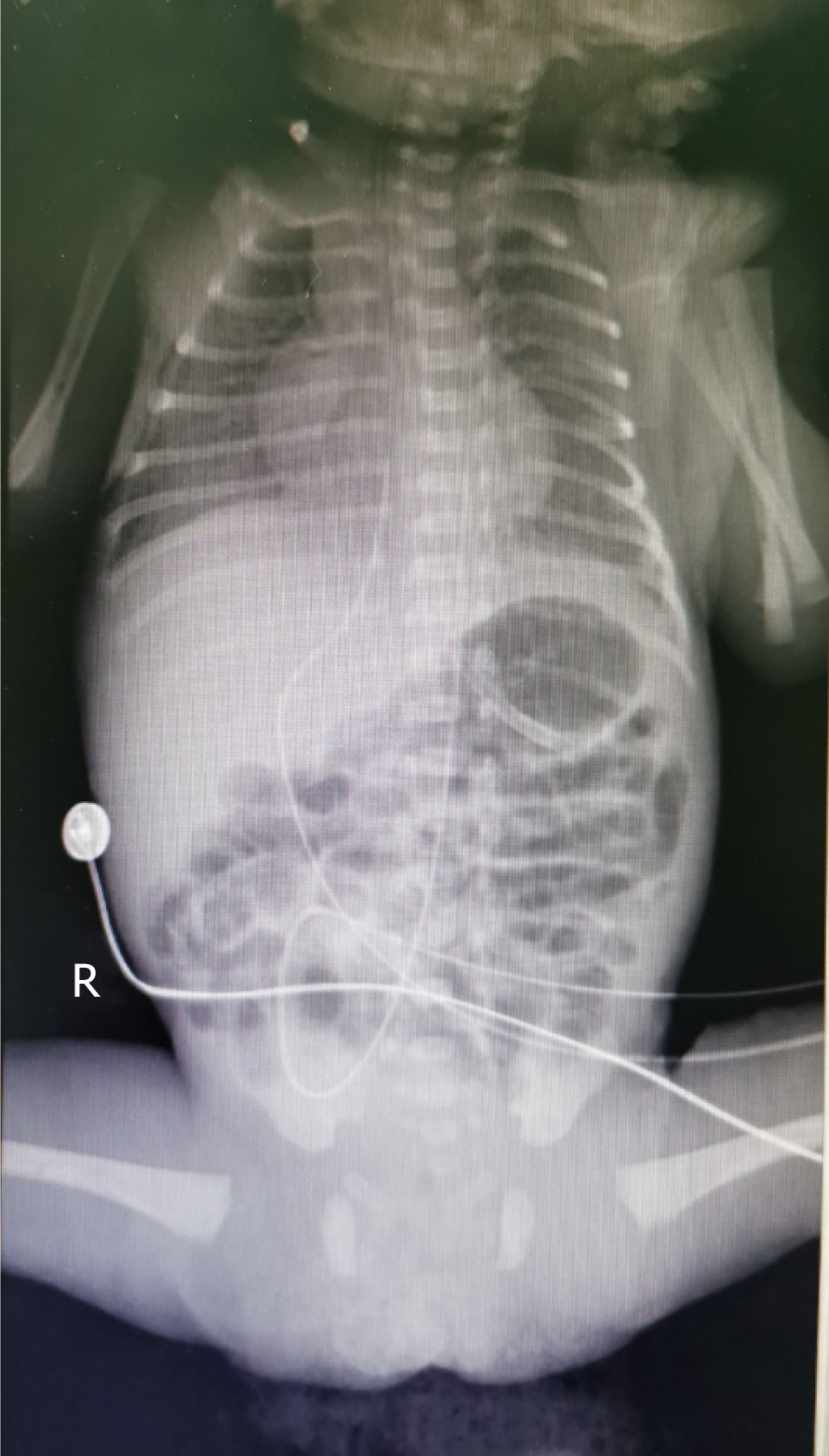
Figure 2 Abdominal X-ray (May 20).
The small intestine showed inflation, but no obvious dilatation of the intestinal lumen or effusion was noted.
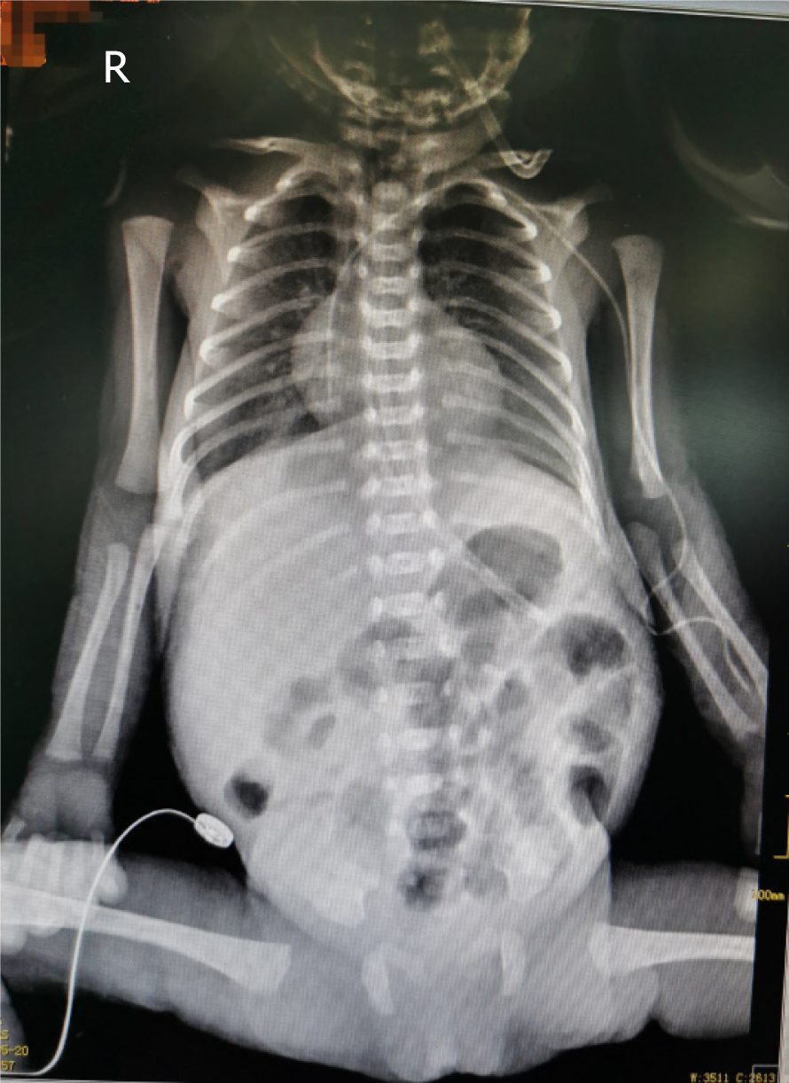
Figure 3 Abdominal X-ray (May 21).
The range of intestinal inflation increased over previous measurements.
- Citation: Huang X, Hu YL, Zhao Y, Chen Q, Li YX. Neonatal necrotizing enterocolitis caused by umbilical arterial catheter-associated abdominal aortic embolism: A case report. World J Clin Cases 2021; 9(22): 6557-6565
- URL: https://www.wjgnet.com/2307-8960/full/v9/i22/6557.htm
- DOI: https://dx.doi.org/10.12998/wjcc.v9.i22.6557


