Copyright
©The Author(s) 2021.
World J Clin Cases. Aug 6, 2021; 9(22): 6552-6556
Published online Aug 6, 2021. doi: 10.12998/wjcc.v9.i22.6552
Published online Aug 6, 2021. doi: 10.12998/wjcc.v9.i22.6552
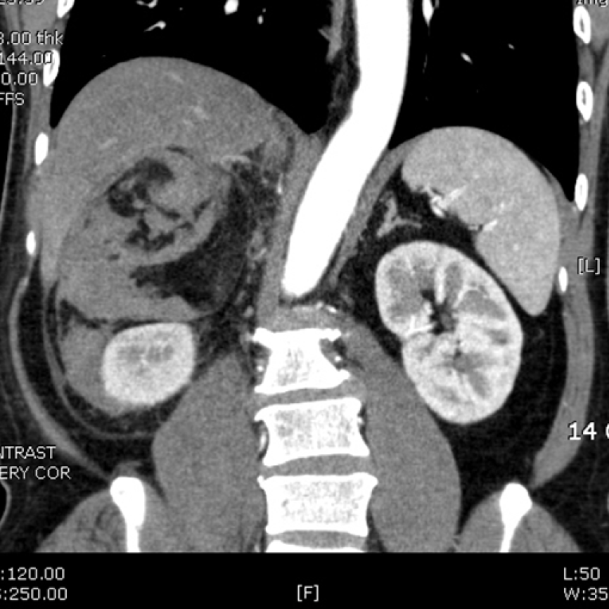
Figure 1 Computed tomography scan of the adrenal myelolipoma.
On computed tomography, adrenal myelolipomas exhibit distinct characteristics, with most of the mass showing fat attenuation. In this case, the tumor was located superior to the right kidney, showing mixed low attenuation due to the fat component and intermediate attenuation because of hemorrhage.
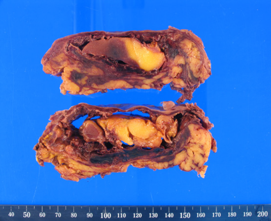
Figure 2 Macroscopic view of the resected tumor.
The tumor consisted of mainly of yellowish fat tissue. However, the capsule was ruptured, and hemorrhage was observed, consistent with the computed tomography scan.
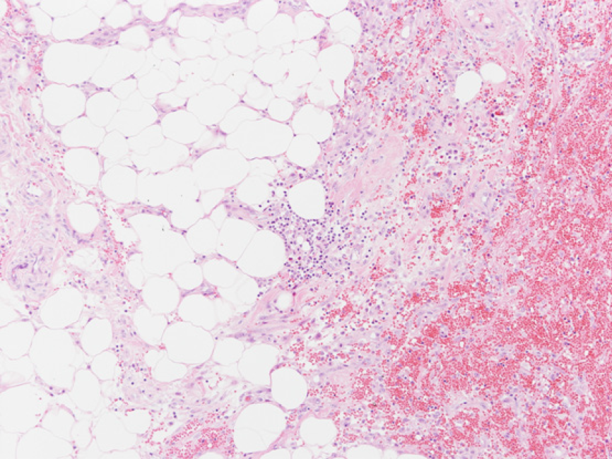
Figure 3 Pathologic findings of the specimen (magnification, × 100).
The specimen showed a mixture of mature adipose tissue and hematopoietic elements.
- Citation: Kim DS, Lee JW, Lee SH. Spontaneous rupture of adrenal myelolipoma as a cause of acute flank pain: A case report. World J Clin Cases 2021; 9(22): 6552-6556
- URL: https://www.wjgnet.com/2307-8960/full/v9/i22/6552.htm
- DOI: https://dx.doi.org/10.12998/wjcc.v9.i22.6552


