Copyright
©The Author(s) 2021.
World J Clin Cases. Aug 6, 2021; 9(22): 6515-6521
Published online Aug 6, 2021. doi: 10.12998/wjcc.v9.i22.6515
Published online Aug 6, 2021. doi: 10.12998/wjcc.v9.i22.6515
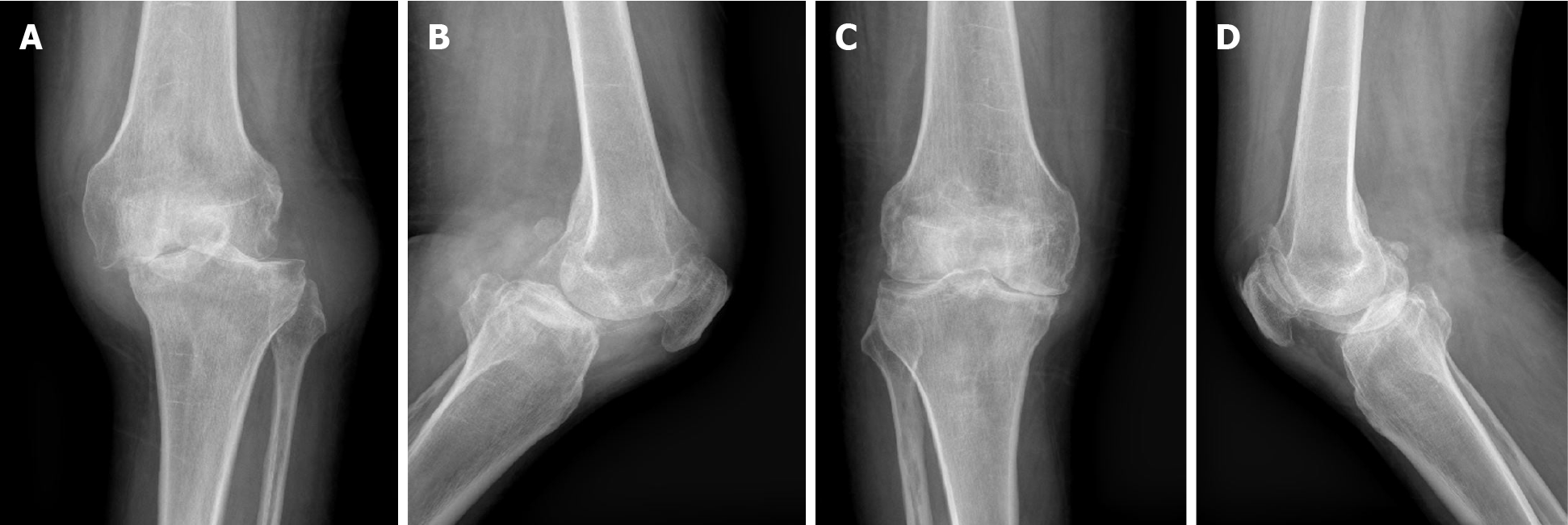
Figure 1 Preoperative X-ray images.
A and B: Left knee front and lateral radiographs; C and D: Right knee front and lateral radiographs.
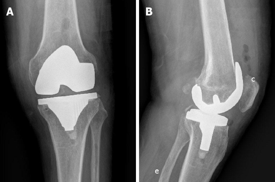
Figure 2 Postoperative front and lateral X-ray images of the left knee.
A: Front X-ray image of the left knee; B: Lateral X-ray image of the left knee.
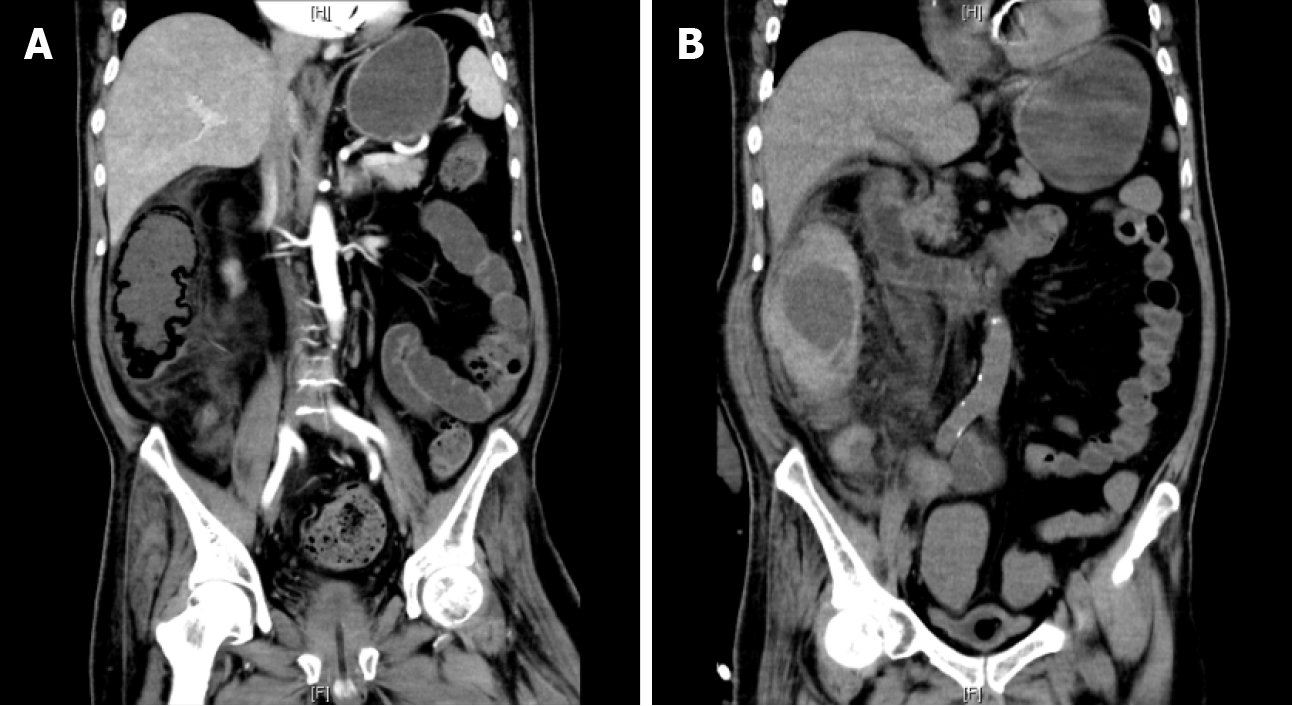
Figure 3 Abdominal computed tomography.
A: The initial abdominal computed tomography (CT) showed ascending colon emphysema with fluid planes and exudative changes; B: The second abdominal CT showed thickening of the wall of the ascending colon with multiple exudative changes around it and possible local hematoma.
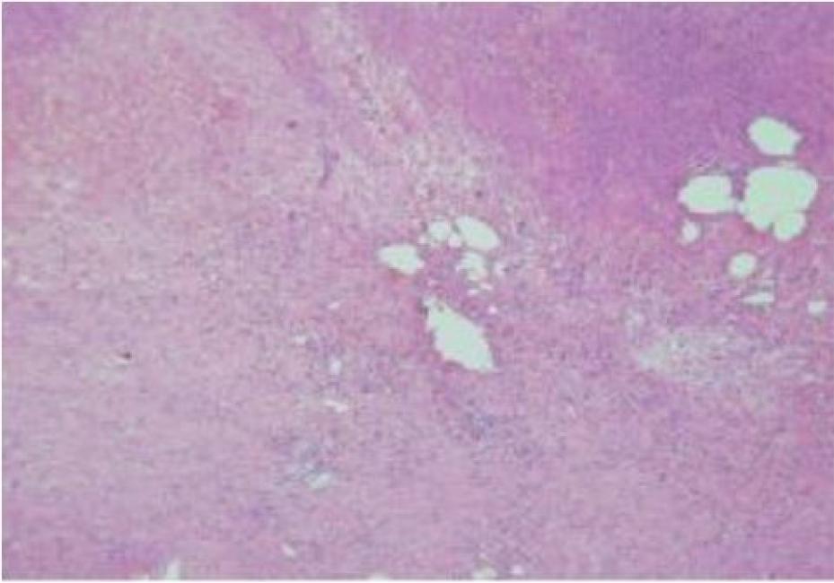
Figure 4 The pathology of the right hemicolectomy specimen showed mucosal surface erosion, inflammatory exudation, and necrosis of the intestinal canal, and the surrounding intestinal wall and mesentery were vasodilated and congested, consistent with ischemic changes of the colon.
- Citation: Zhang SY, He BJ, Xu HH, Xiao MM, Zhang JJ, Tong PJ, Mao Q. Concealed mesenteric ischemia after total knee arthroplasty: A case report. World J Clin Cases 2021; 9(22): 6515-6521
- URL: https://www.wjgnet.com/2307-8960/full/v9/i22/6515.htm
- DOI: https://dx.doi.org/10.12998/wjcc.v9.i22.6515


