Copyright
©The Author(s) 2021.
World J Clin Cases. Aug 6, 2021; 9(22): 6493-6500
Published online Aug 6, 2021. doi: 10.12998/wjcc.v9.i22.6493
Published online Aug 6, 2021. doi: 10.12998/wjcc.v9.i22.6493
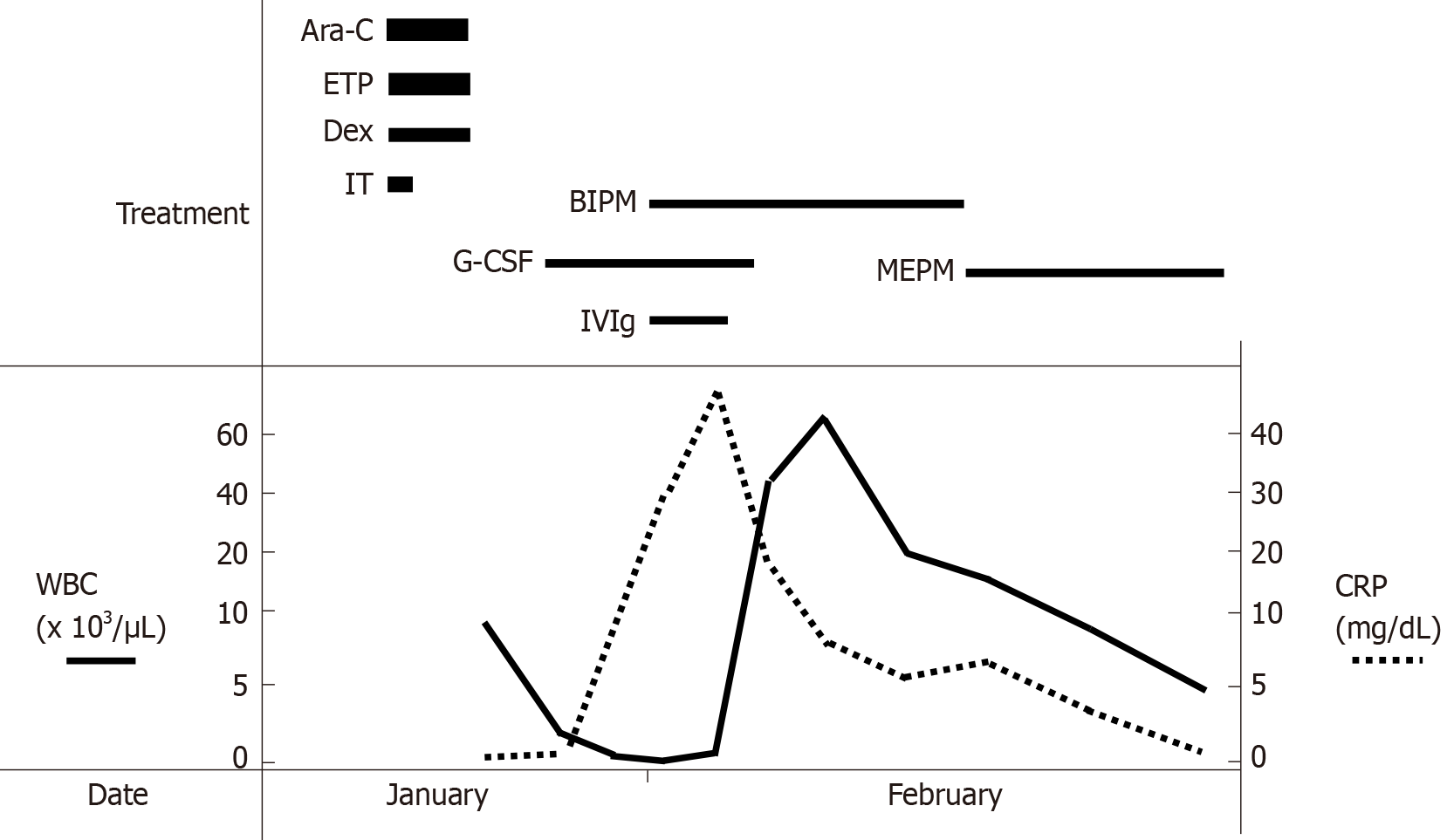
Figure 1 Clinical course of this patient.
Ara-C: Cytarabine; ETP: Etoposide; Dex: Dexamethasone; IT: Intrathecal injection; G-CSF: Granulocyte-colony stimulating factor; BIPM: Biapenem; MEPM: Meropenem; IVIg: Intra-venous immunoglobulin; WBC: White blood cell count; CRP: C-reactive protein.
Figure 2 Abdominal computed tomography (day 11).
Marked thickening of the gastric wall (orange arrow), suggestive of hepatic portal venous gas (white arrow), and low-density areas (blue arrow) were found.
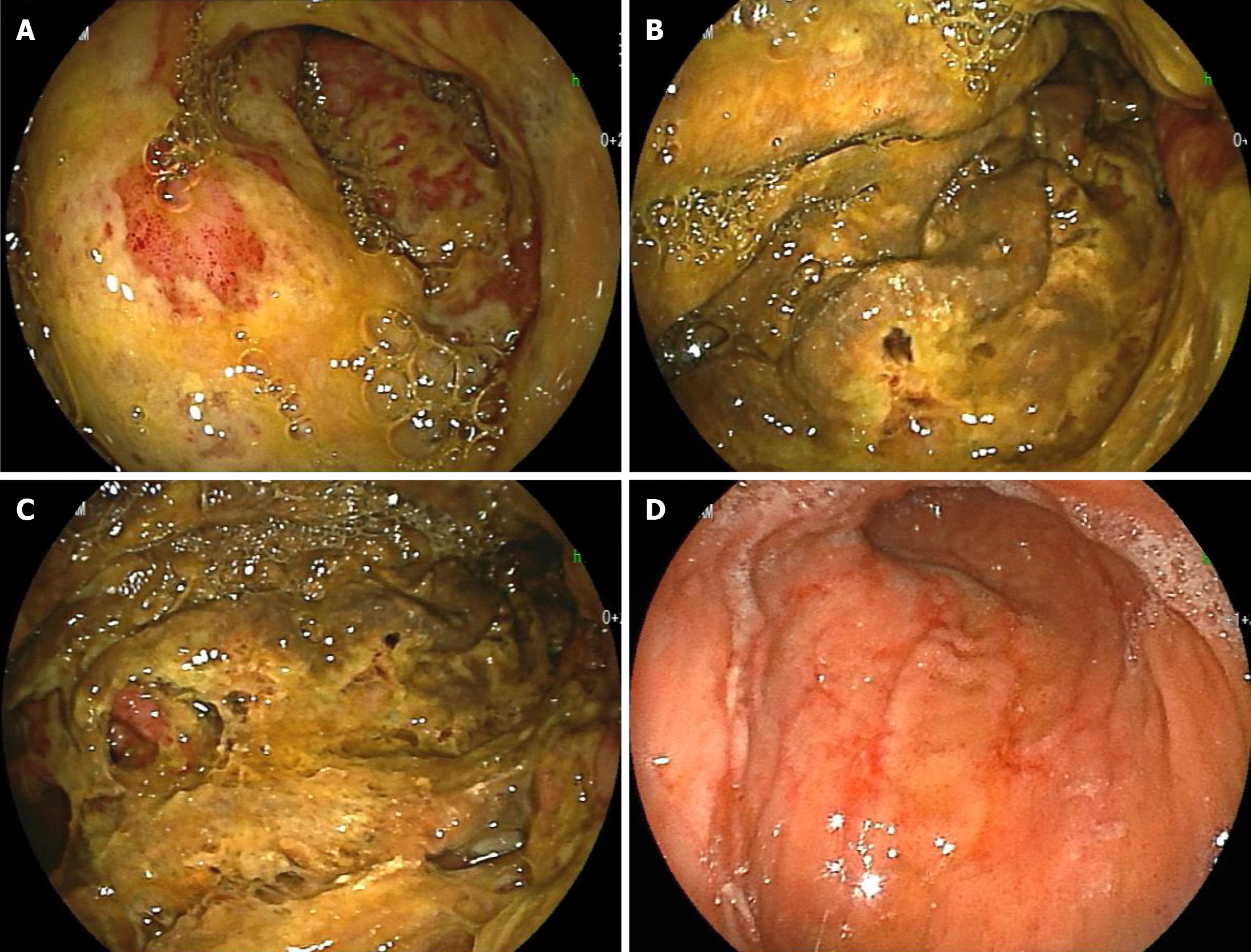
Figure 3 Esophagogastroduodenoscopy.
A-C: Marked thickening of the gastric wall in the corpus of the stomach and yellow-green pseudomembrane-like tissue covering the superficial mucosa were observed (day 14); D: The above abnormal findings were improved, and linear redness, erosion and ulcerative mucosal changes were observed (day 29).
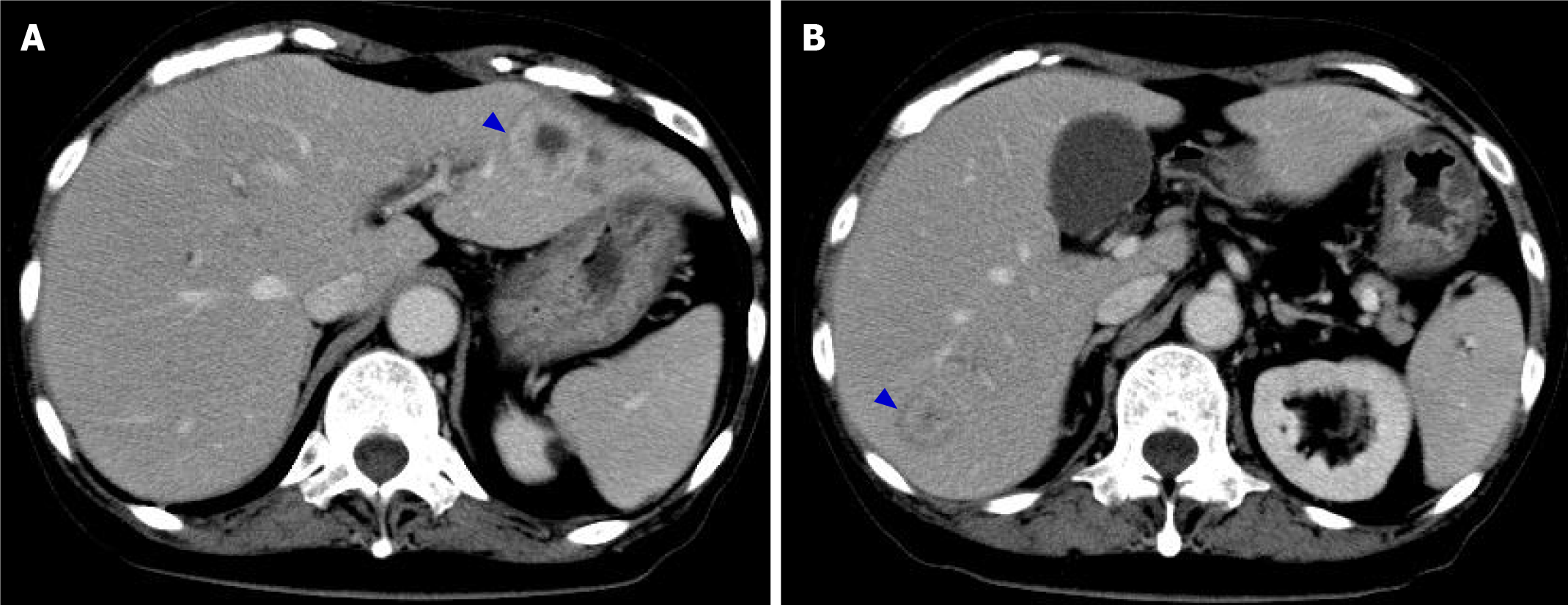
Figure 4 Abdominal computed tomography (day 29).
The thickening of the gastric wall was improved, and signs suggestive of hepatic portal venous gas disappeared. The low-density areas in liver S3 and S7 observed 18 d ago were changed the findings consistent with abscesses (blue arrow).
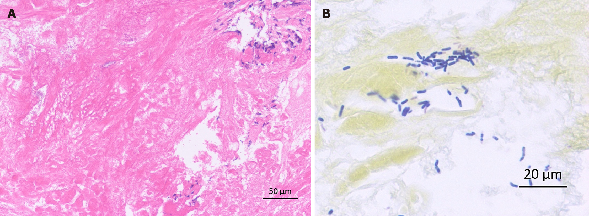
Figure 5 Histopathology of the gastric biopsy.
A: Mostly necrotic tissue and partly infiltration by lymphocytes were observed (hematoxylin & eosin stain); B: Bacilli were gram-positive (gram stain).
- Citation: Saito M, Morioka M, Izumiyama K, Mori A, Ogasawara R, Kondo T, Miyajima T, Yokoyama E, Tanikawa S. Phlegmonous gastritis developed during chemotherapy for acute lymphocytic leukemia: A case report. World J Clin Cases 2021; 9(22): 6493-6500
- URL: https://www.wjgnet.com/2307-8960/full/v9/i22/6493.htm
- DOI: https://dx.doi.org/10.12998/wjcc.v9.i22.6493


