Copyright
©The Author(s) 2021.
World J Clin Cases. Aug 6, 2021; 9(22): 6469-6477
Published online Aug 6, 2021. doi: 10.12998/wjcc.v9.i22.6469
Published online Aug 6, 2021. doi: 10.12998/wjcc.v9.i22.6469
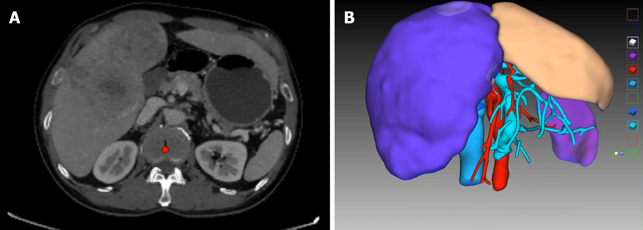
Figure 1 Computed tomography result and three-dimensional reconstruction of the patient at first presentation.
A: Venous phase contrast-enhanced computed tomography shows huge space-occupying lesions in the left medial lobe and right liver, and the left lateral lobe is normal; B: Virtual resection on the three-dimensional liver model.
Figure 2 Conversion therapy.
A: Embolization of the right portal vein; B: Chemoembolization of the tumor in left medial lobe via left hepatic artery and abdominal trunk; C: Chemoembolization of tumors in the right liver via right hepatic artery and superior mesenteric artery.
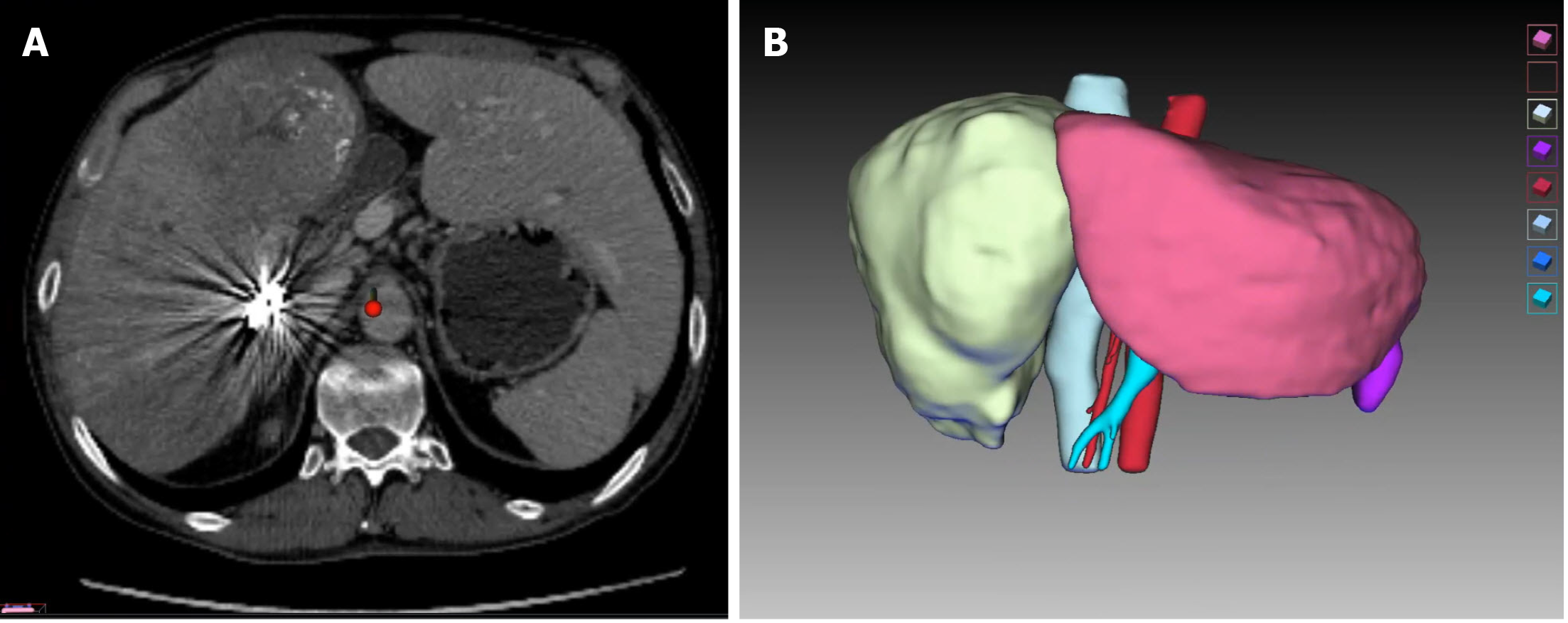
Figure 3 Computed tomography result and three-dimensional reconstruction of the patient prior to surgery.
A: Pre-operative venous phase contrast-enhanced computed tomography shows huge space-occupying lesions in the left medial lobe and right liver and obvious hyperplasia in the left lateral lobe; B: Virtual resection on the three-dimensional liver model.
Figure 4 Intraoperative observation.
A: Liver remnant of the left lateral lobe, with normal color; B: Liver cutting surface, with intact Glisson sheath covering the left lateral lobe.
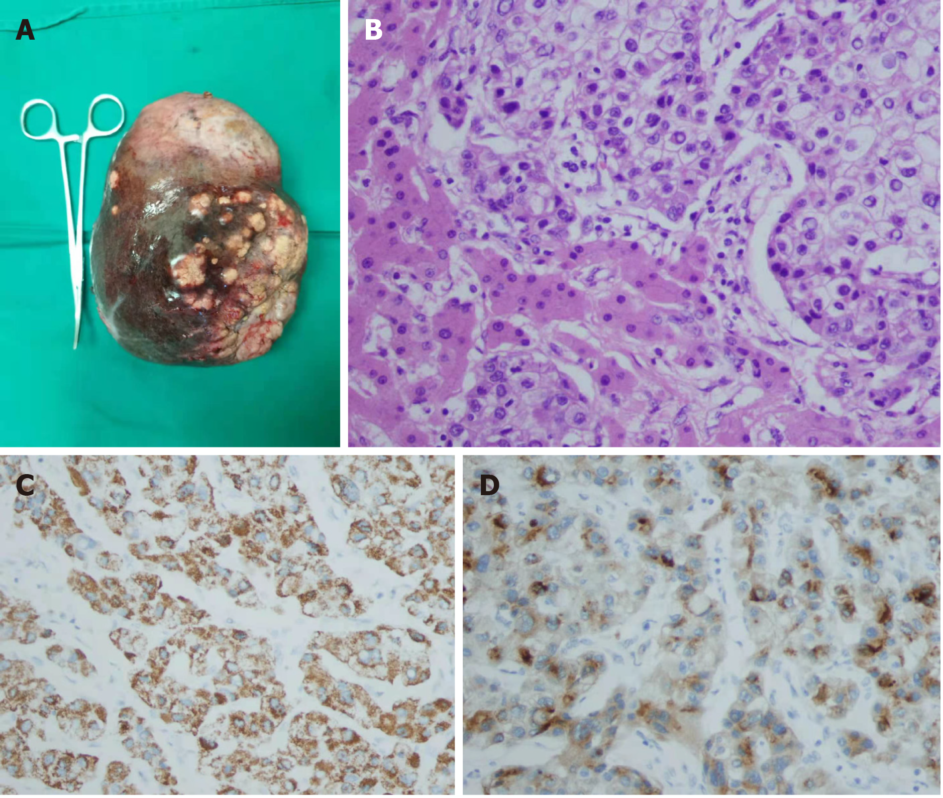
Figure 5 Resected specimen (diaphragmatic surface of liver).
A: Microscopic findings. Hematoxylin and eosin (H&E) staining showing polygonal tumor cells in a trabecular arrangement, H&E × 400; B-D: Immunohistochemistry showing a strong and diffuse cytoplasmic (B) positivity with Hep Par 1 (C) and glypican 3 (D), × 400. The tumor cells were negative for cytokeratin 7 and cytokeratin 20.
Figure 6 Postoperative recovery and short-term follow-up.
A: Abdominal plain computed tomography (CT) at discharge showed no accumulation of fluid in the abdominal cavity, whereas there was hyperplasia of the left lateral lobe; B: Abdominal incision at discharge; C: A recent CT scan showed that there was no tumor recurrence, and the hyperplasia of the left lateral lobe was obvious.
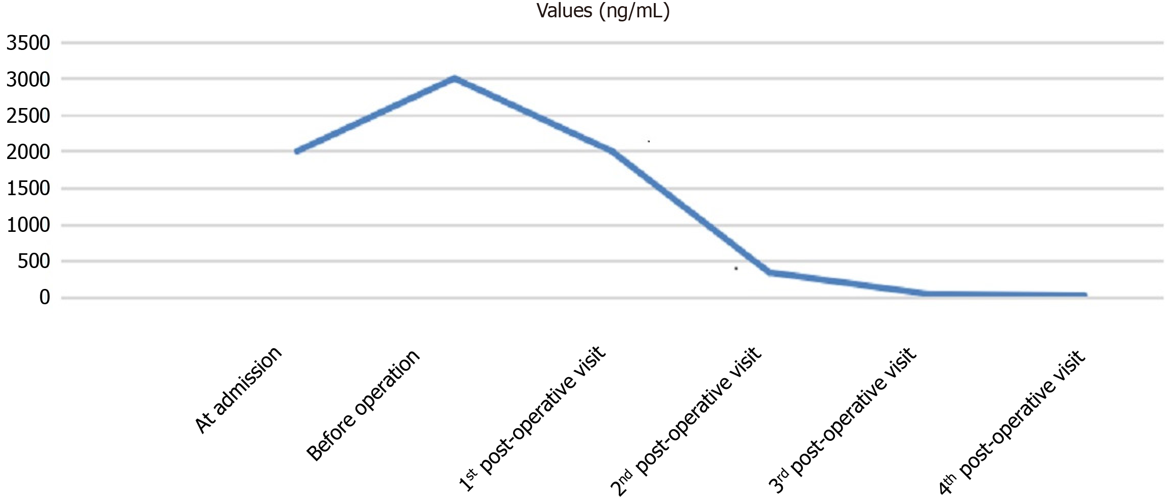
Figure 7 Change in alpha-fetal protein level.
- Citation: Zhang JJ, Wang ZX, Niu JX, Zhang M, An N, Li PF, Zheng WH. Successful totally laparoscopic right trihepatectomy following conversion therapy for hepatocellular carcinoma: A case report. World J Clin Cases 2021; 9(22): 6469-6477
- URL: https://www.wjgnet.com/2307-8960/full/v9/i22/6469.htm
- DOI: https://dx.doi.org/10.12998/wjcc.v9.i22.6469


