Copyright
©The Author(s) 2021.
World J Clin Cases. Aug 6, 2021; 9(22): 6443-6449
Published online Aug 6, 2021. doi: 10.12998/wjcc.v9.i22.6443
Published online Aug 6, 2021. doi: 10.12998/wjcc.v9.i22.6443
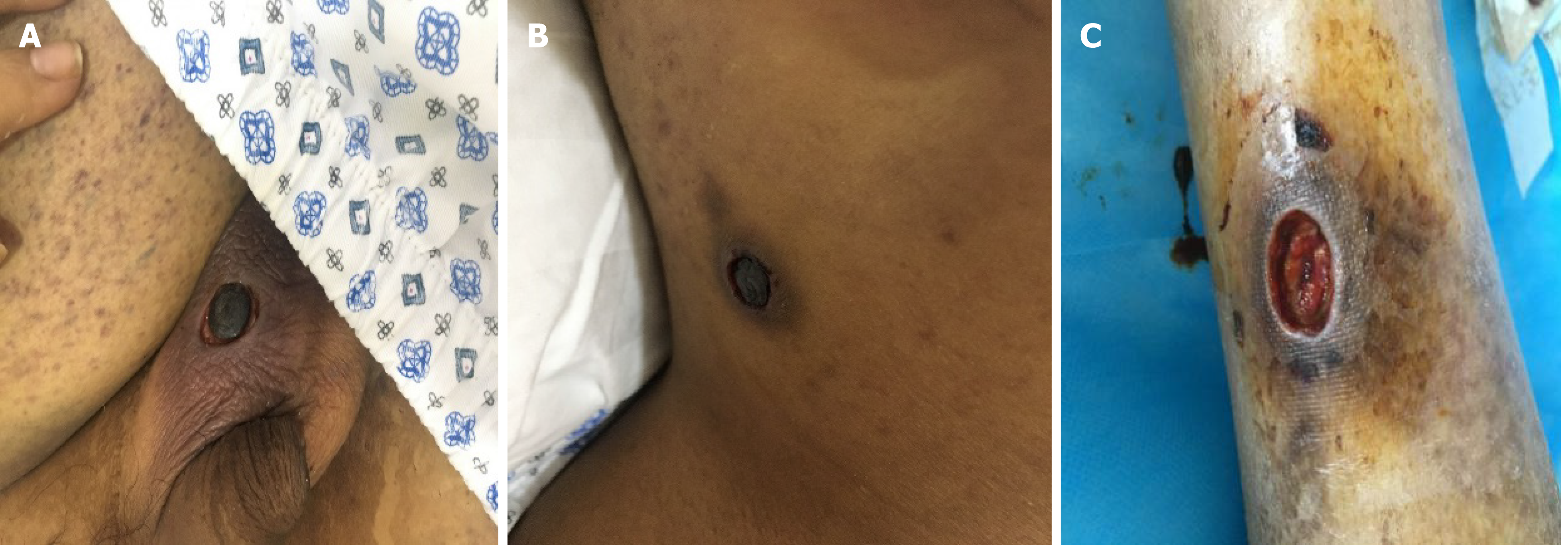
Figure 1 Images of the nodules.
A: Left scrotum; B: Right neck; C: Right lower leg. Painful nodules are seen.
Figure 2 Staining images.
A: Hematoxylin and eosin staining; B: Methenamine sliver staining; C: Periodic acid–Schiff staining. Fusarium hyphae and spores are seen in the right neck lesion (magnification × 400).
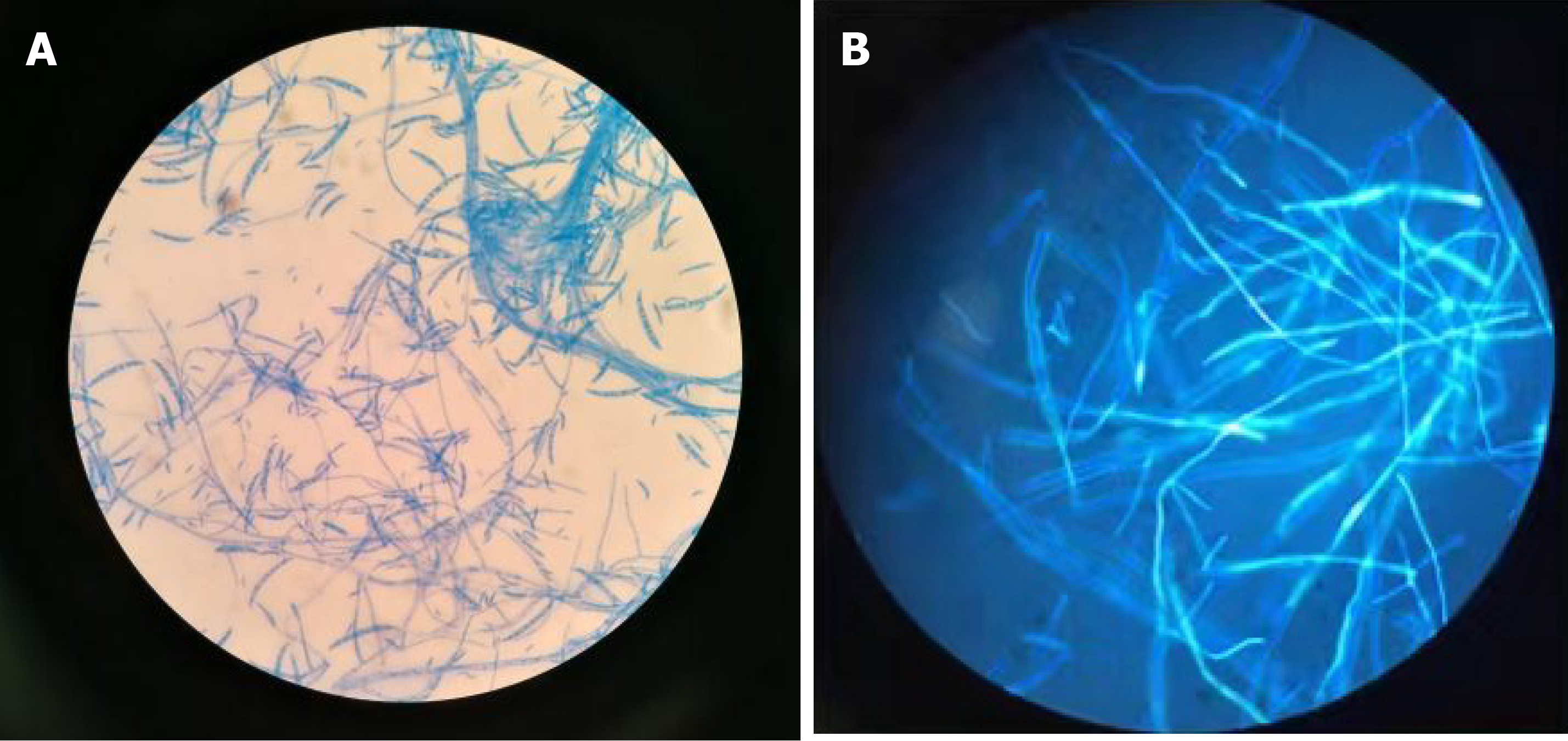
Figure 3 Microscopic examination of fungus.
A: Ordinary microscope; B: Fluorescence microscopy.
Figure 4 Images of the lesions after treatment.
A: Left scrotum; B: Right neck; C: Right lower leg. The skin lesions have gradually subsided.
- Citation: Yao YF, Feng J, Liu J, Chen CF, Yu B, Hu XP. Disseminated infection by Fusarium solani in acute lymphocytic leukemia: A case report. World J Clin Cases 2021; 9(22): 6443-6449
- URL: https://www.wjgnet.com/2307-8960/full/v9/i22/6443.htm
- DOI: https://dx.doi.org/10.12998/wjcc.v9.i22.6443


