Copyright
©The Author(s) 2021.
World J Clin Cases. Aug 6, 2021; 9(22): 6410-6417
Published online Aug 6, 2021. doi: 10.12998/wjcc.v9.i22.6410
Published online Aug 6, 2021. doi: 10.12998/wjcc.v9.i22.6410
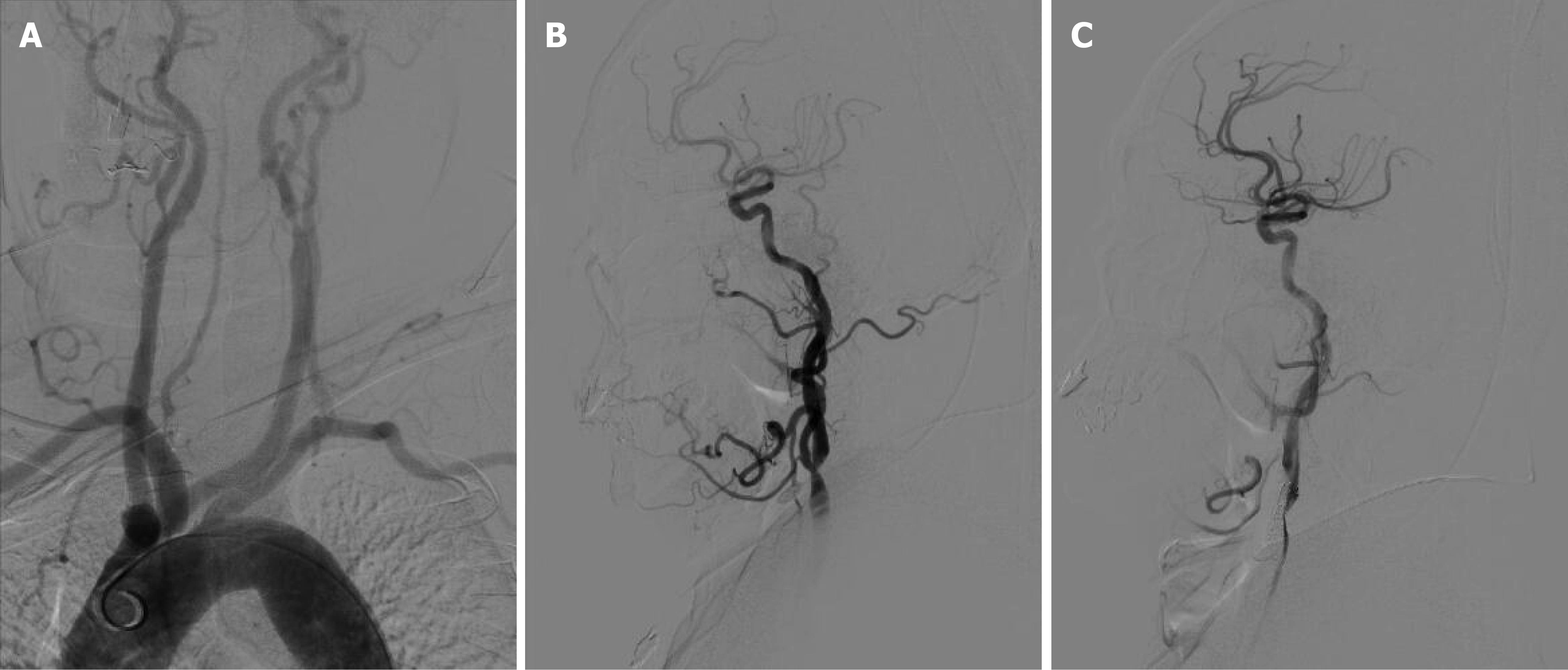
Figure 1 Preoperative digital subtraction angiography findings.
A: The type III aortic arch; B: The bifurcation of the left common carotid artery was severely stenosed; C: The stenosis at the bifurcation of the left common carotid artery was significantly improved.
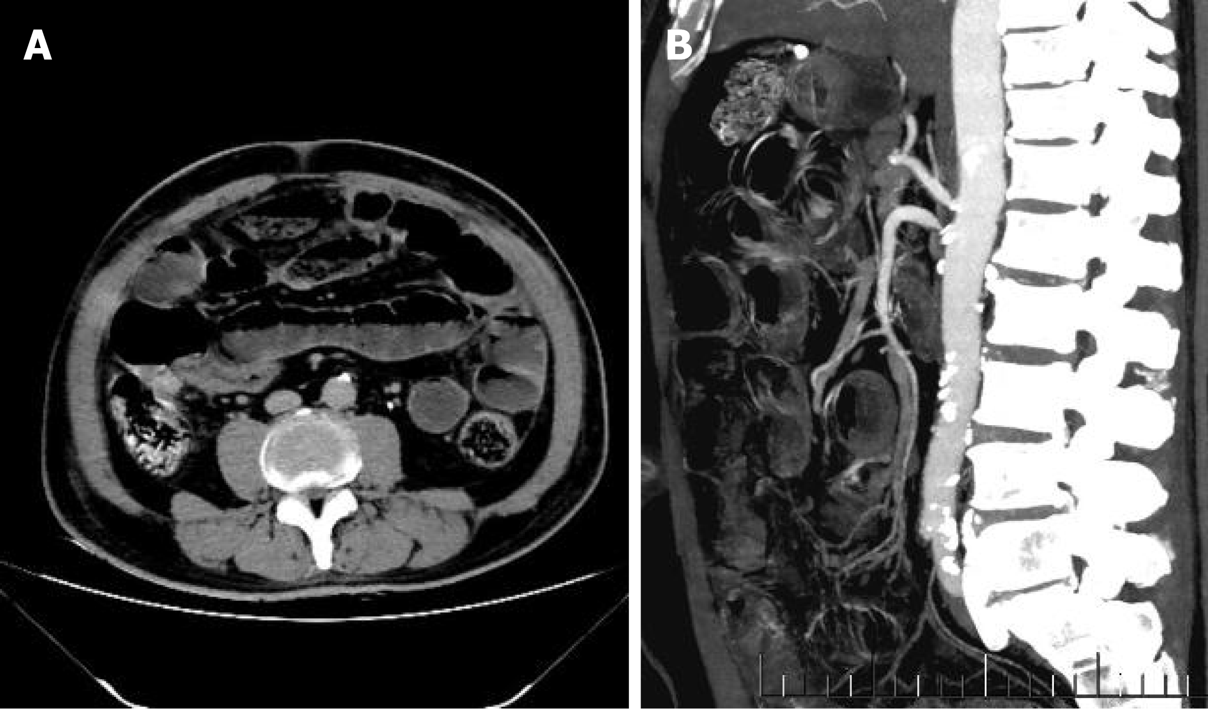
Figure 2 Abdominal enhanced computed tomography changes after carotid artery stenting.
A: Intestinal obstruction and edema of the intestinal wall; B: The superior mesenteric artery showed severe stenosis and poor distal angiography.
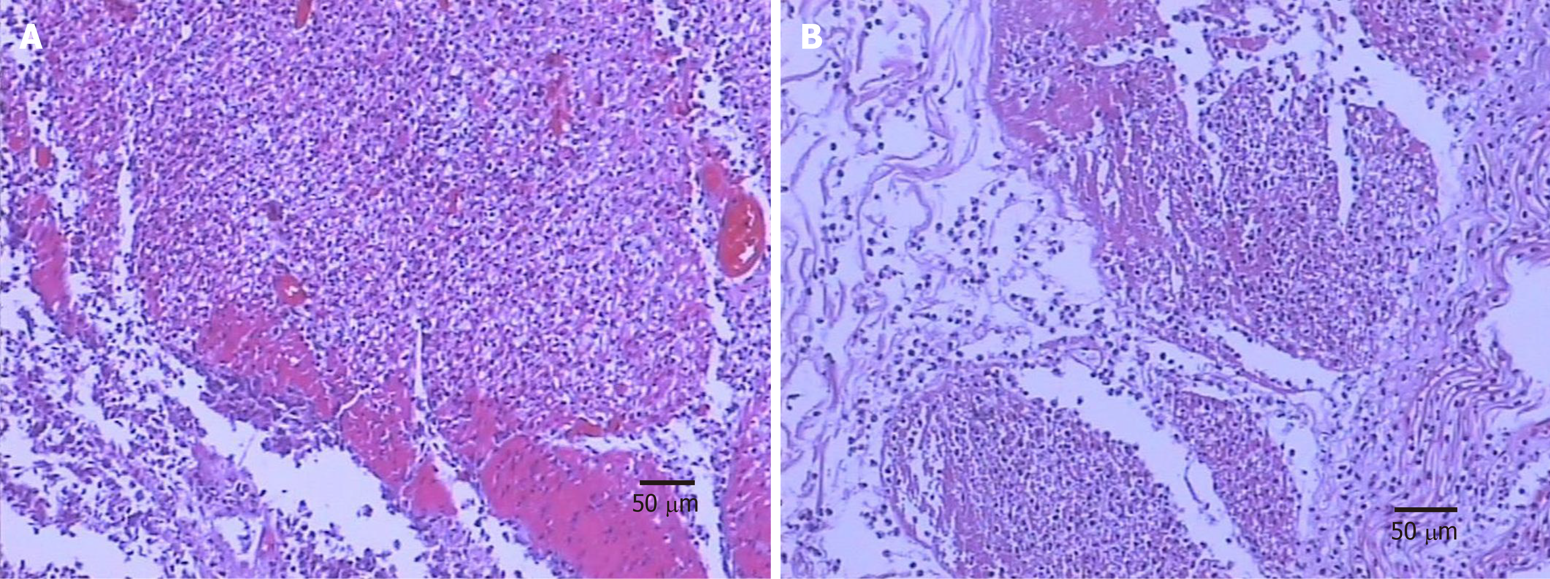
Figure 3 Pathology of the ileectomy site after partial ileectomy.
A and B: Intestinal mucosa necrosis, exfoliation, full-thickness vasodilation and hyperemia, fibrinous exudation, and necrosis of serous membrane are visible, thus indicating ileal hemorrhagic infarction.
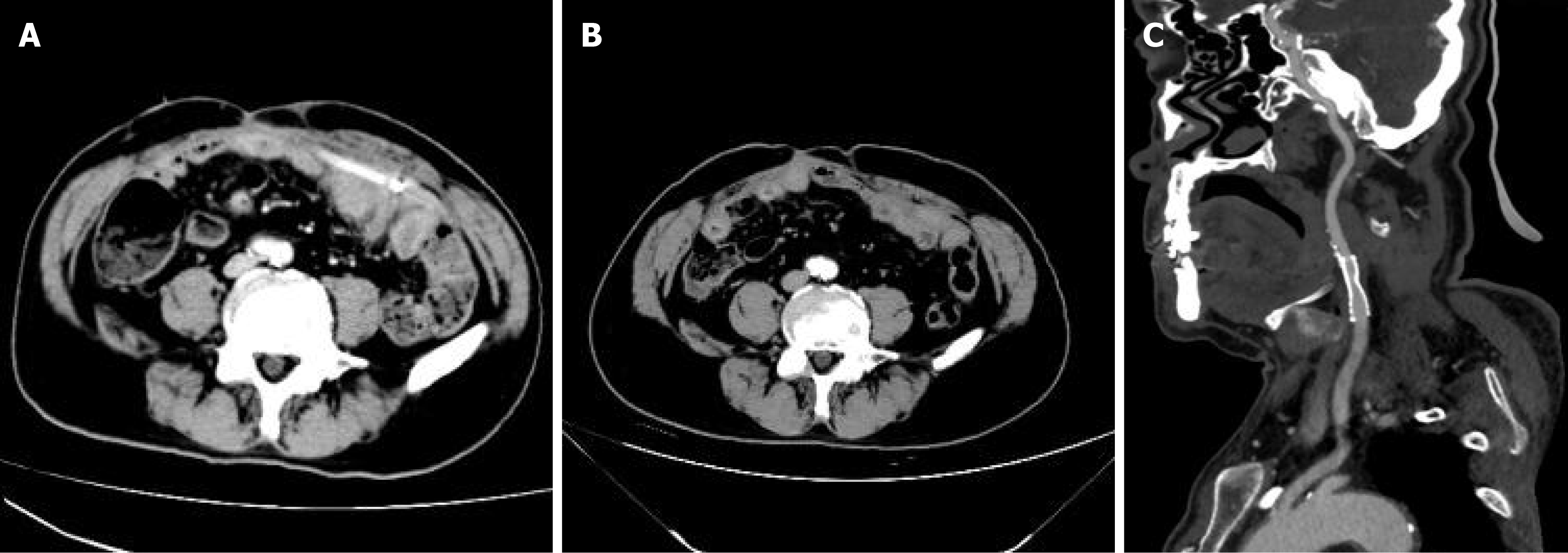
Figure 4 Abdominal enhanced computed tomography changes after the abdominal drainage tube under the guidance of ultrasound.
A: The drainage tube was unobstructed without intestinal obstruction; B: Abdominal enhanced computed tomography after removal of the abdominal drainage tube showed no intestinal obstruction or flatulence; C: Follow-up computed tomography angiography at the 6-mo after carotid artery stenting showed that the carotid artery stent was patent.
- Citation: Xu XY, Shen W, Li G, Wang XF, Xu Y. Ileal hemorrhagic infarction after carotid artery stenting: A case report and review of the literature. World J Clin Cases 2021; 9(22): 6410-6417
- URL: https://www.wjgnet.com/2307-8960/full/v9/i22/6410.htm
- DOI: https://dx.doi.org/10.12998/wjcc.v9.i22.6410


