Copyright
©The Author(s) 2021.
World J Clin Cases. Jul 26, 2021; 9(21): 6138-6144
Published online Jul 26, 2021. doi: 10.12998/wjcc.v9.i21.6138
Published online Jul 26, 2021. doi: 10.12998/wjcc.v9.i21.6138
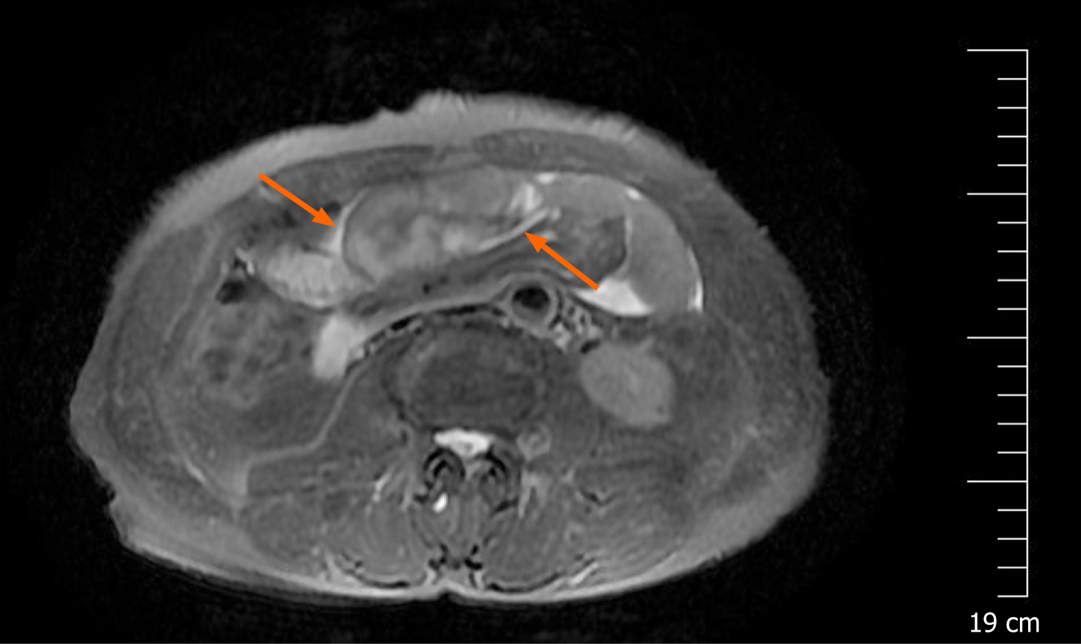
Figure 1 T2 weighted magnetic resonance imaging in axial plane showed bowel loops clustered in a cocoon-like shape that were encased by a thick membrane (arrows).
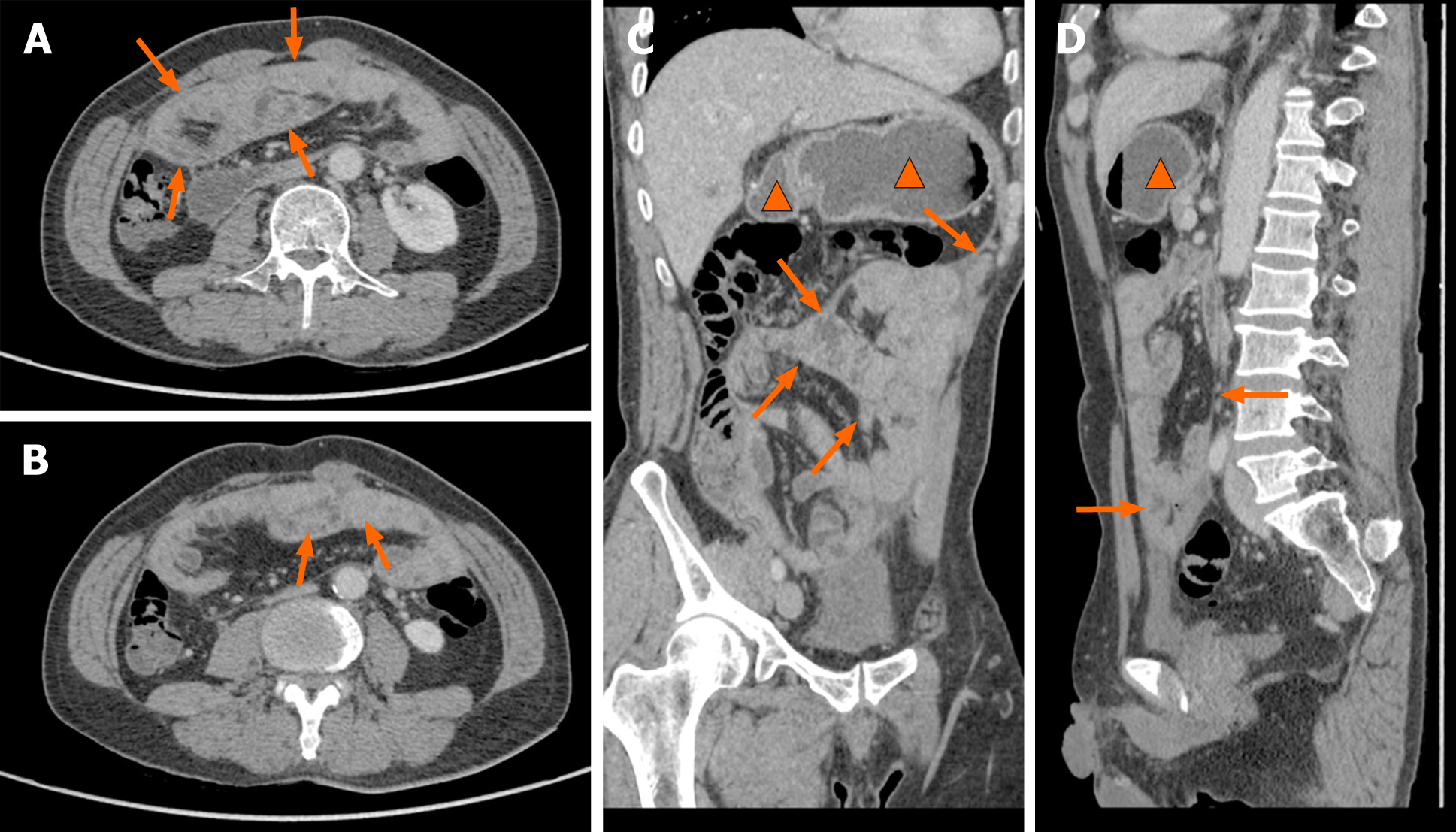
Figure 2 Abdominal contrast-enhanced computed tomography.
A: A thin, membrane-like sac encapsulating the small intestine in an abdominal contrast-enhanced computed tomography image (arrows); B: A thin, membrane-like sac encapsulating the small intestine from a different plane (arrows); C: The dilated gastric cavity (triangles) and the duodenal lumen (arrows) with fluid retention in the sagittal plane in an abdominal contrast-enhanced computed tomography image; D: The dilated gastric cavity (triangles) and the duodenal lumen (arrows) with fluid retention.
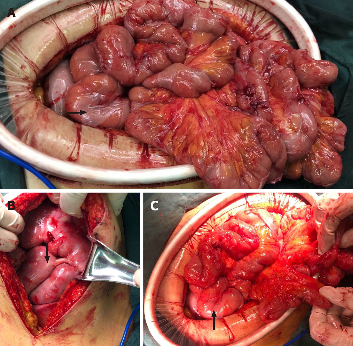
Figure 3 Intraoperative findings.
A: A broken white fibrous membrane encasing loops of the small intestine and the greater omentum (arrow); B: An intact fibrous membrane after laparotomy (arrow); C: A broken membrane encasing loops of the small intestine (arrow).
Figure 4 Comparison of contrast-enhanced computed tomography images before and after surgery.
A: A thin, membrane-like sac before laparoscopic surgery (arrows); B: The membrane was dissected and the trapped organs were released.
- Citation: Yin MY, Qian LJ, Xi LT, Yu YX, Shi YQ, Liu L, Xu CF. Encapsulating peritoneal sclerosis in an AMA-M2 positive patient: A case report. World J Clin Cases 2021; 9(21): 6138-6144
- URL: https://www.wjgnet.com/2307-8960/full/v9/i21/6138.htm
- DOI: https://dx.doi.org/10.12998/wjcc.v9.i21.6138


