Copyright
©The Author(s) 2021.
World J Clin Cases. Jul 26, 2021; 9(21): 6110-6124
Published online Jul 26, 2021. doi: 10.12998/wjcc.v9.i21.6110
Published online Jul 26, 2021. doi: 10.12998/wjcc.v9.i21.6110
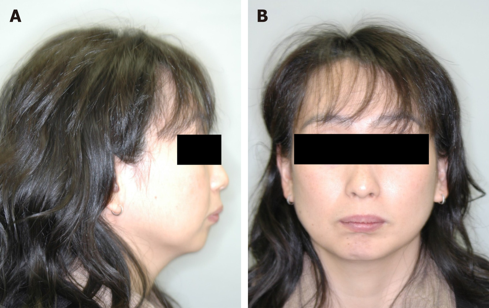
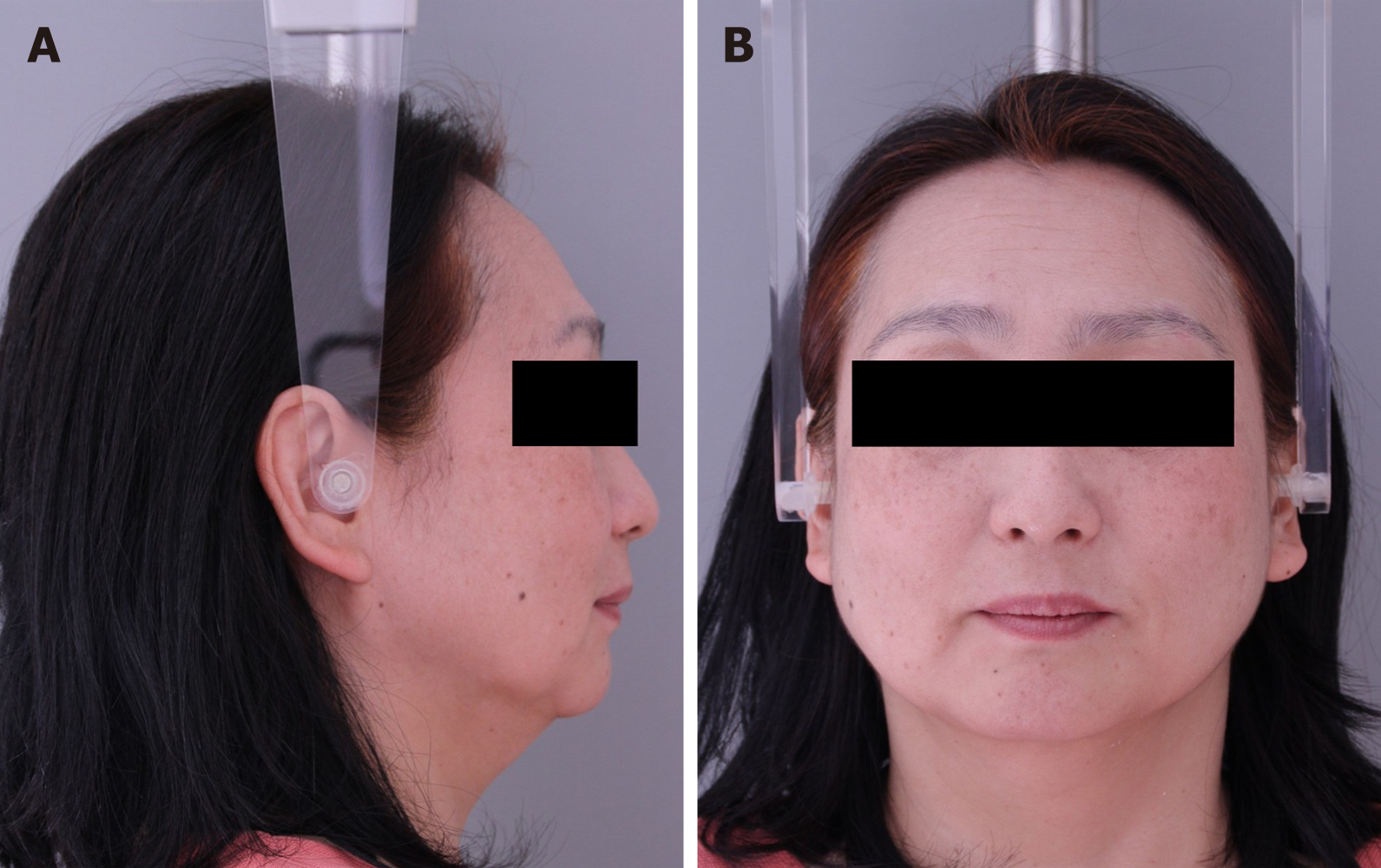
Figure 1 Pre-treatment facial photographs.
A: Lateral view; B: Front view.
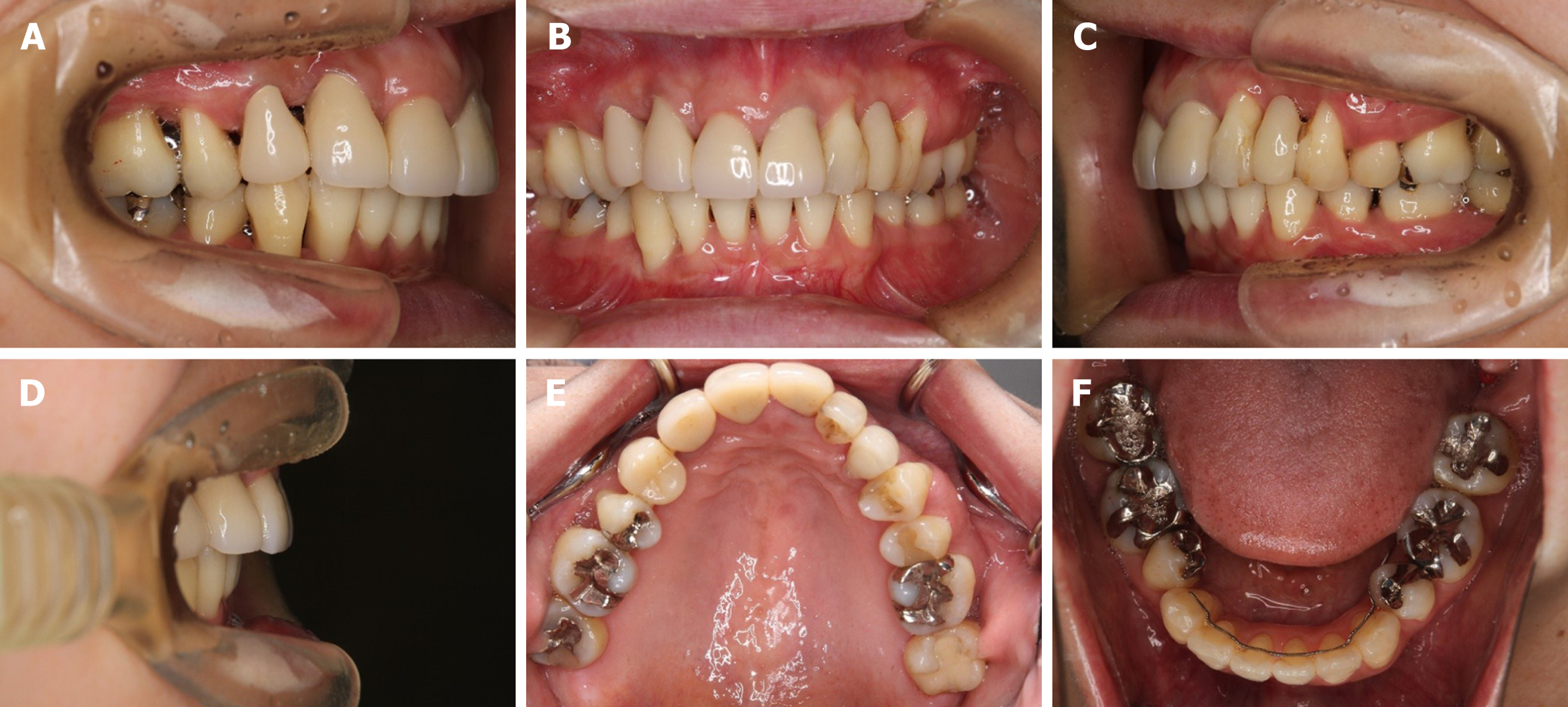
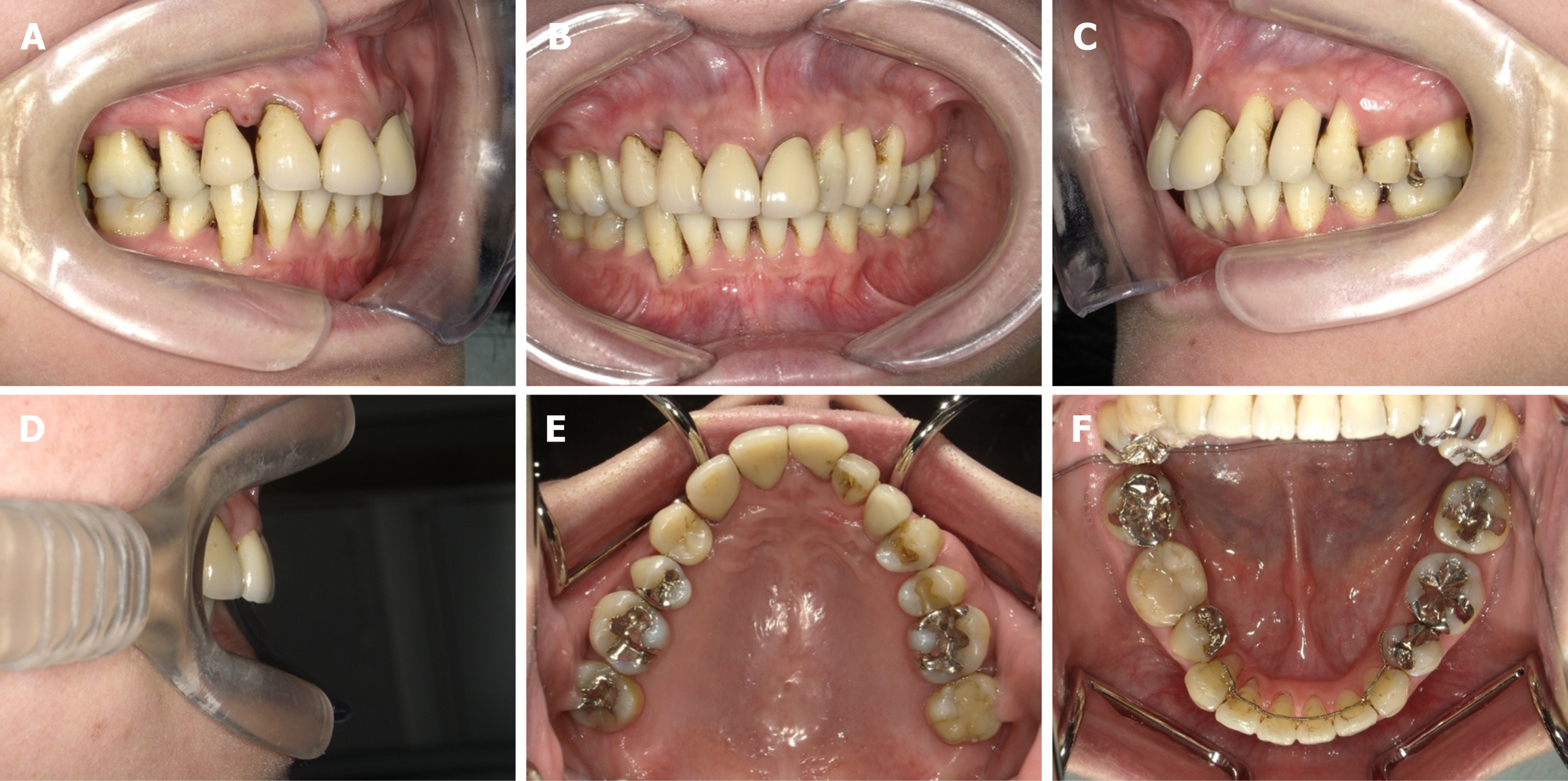
Figure 2 Pre-treatment intraoral photographs.
A: Right side view; B: Front view; C: Left side view; D: Incisal view; E: Upper occlusal view; F: Lower occlusal view.
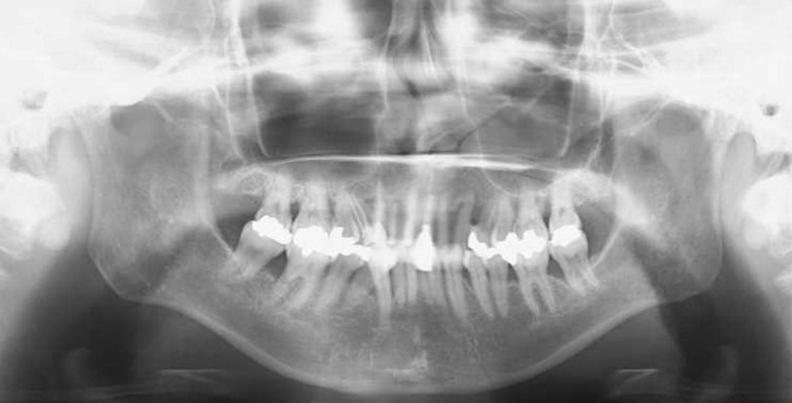
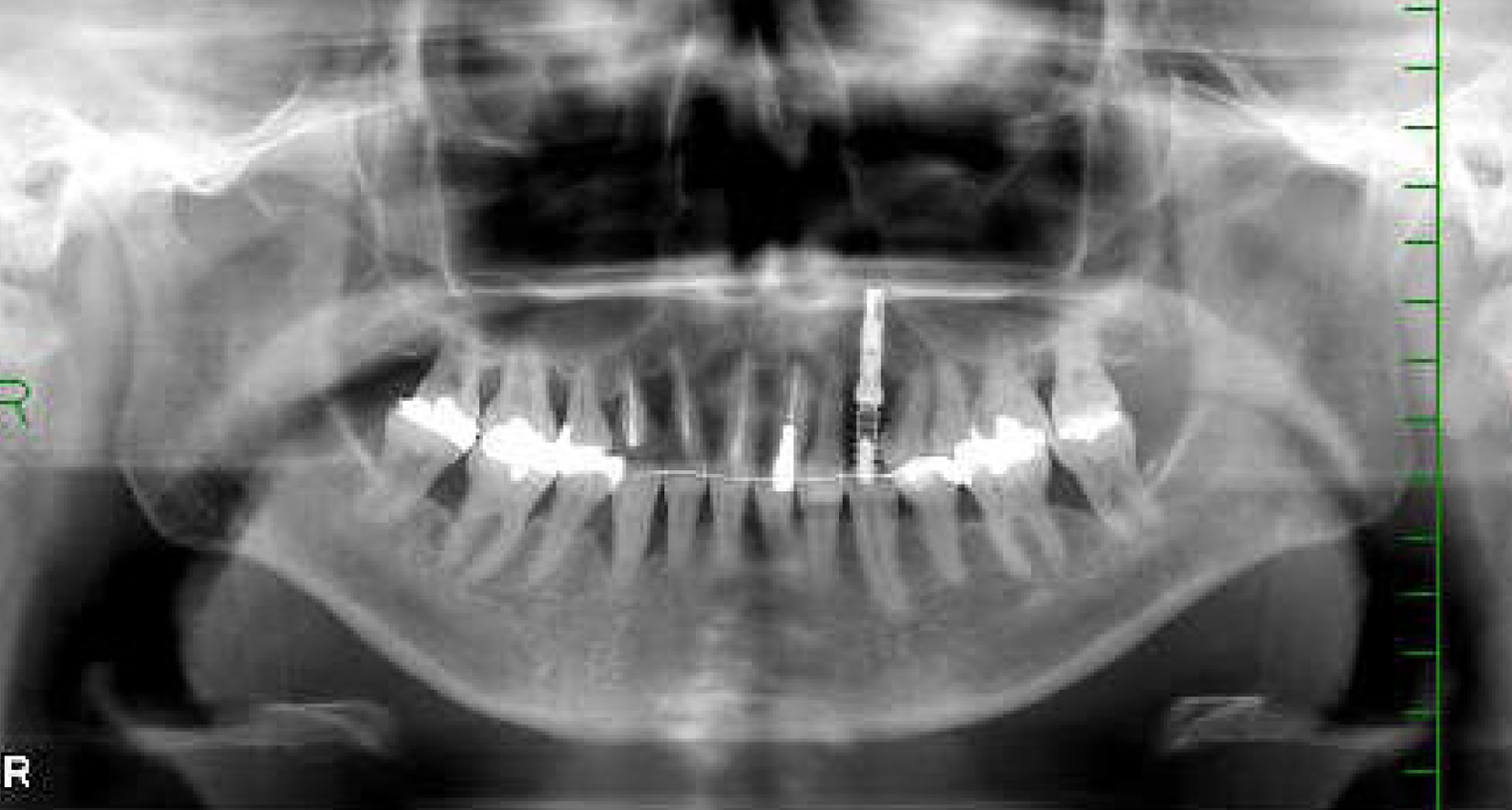
Figure 3 Pre-treatment panoramic radiograph.
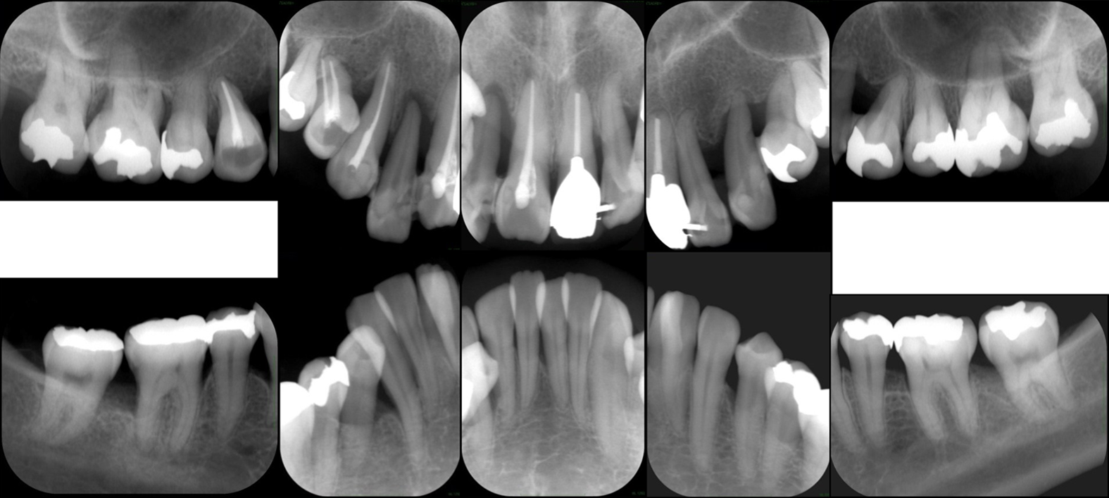
Figure 4 Pre-treatment full-mouth set of dental radiographs.
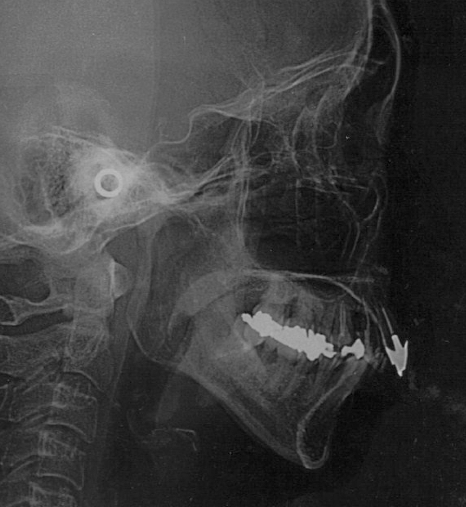
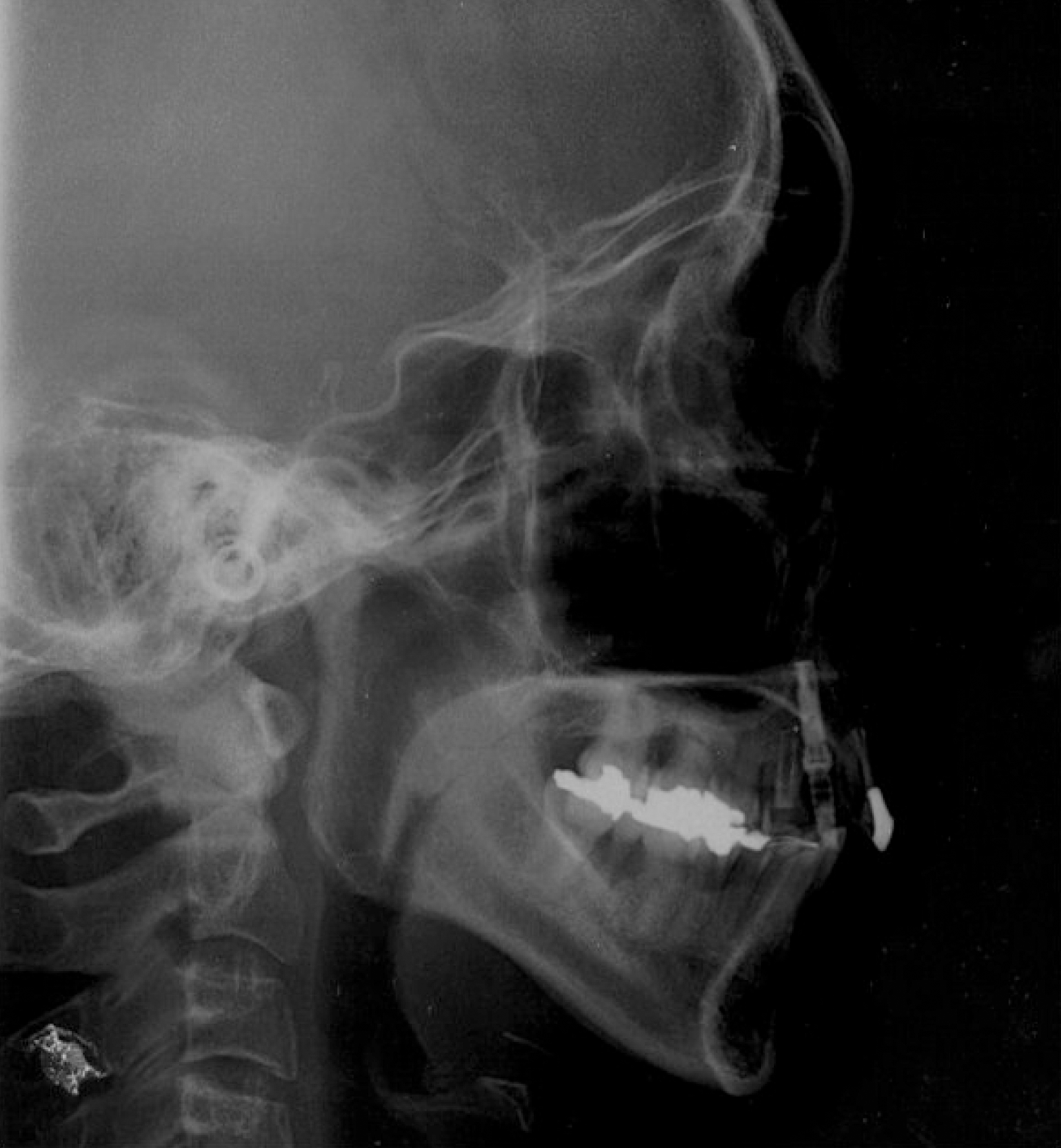
Figure 5 Pre-treatment cephalometric radiograph.
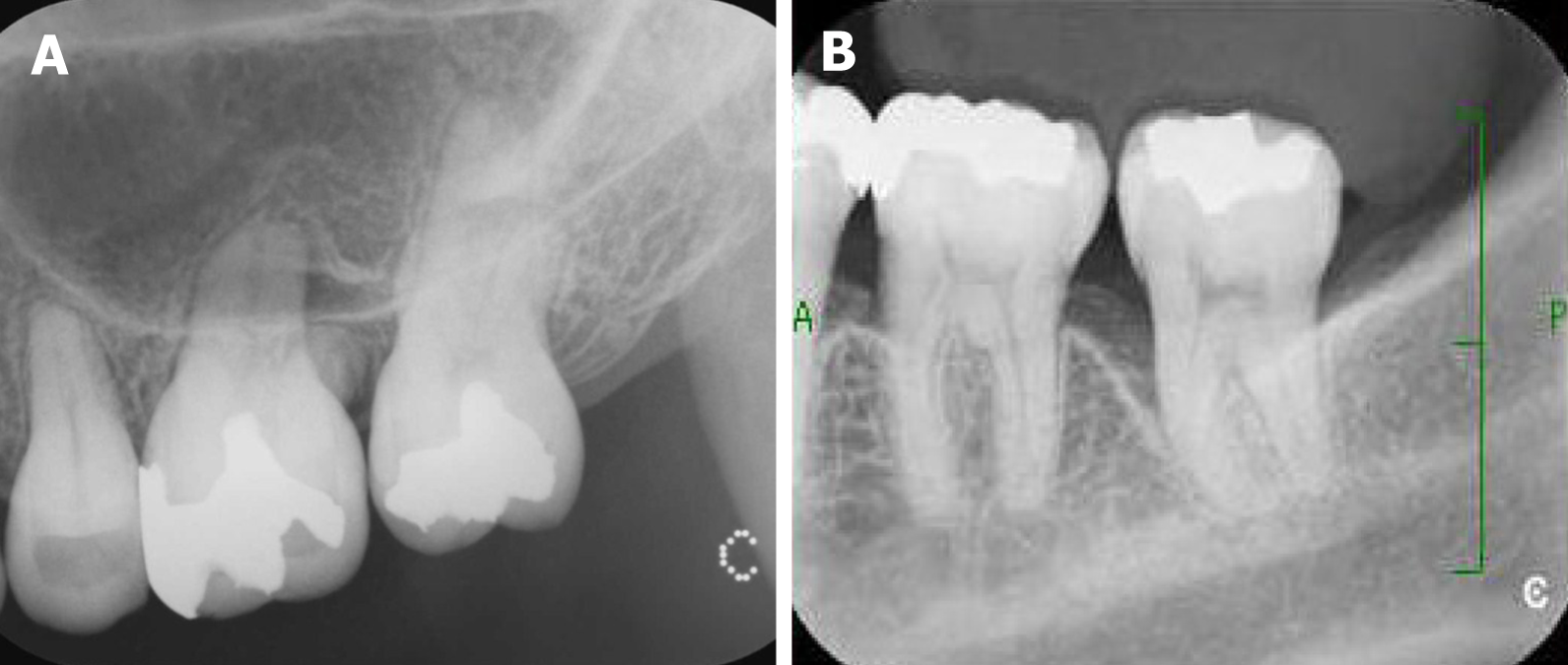
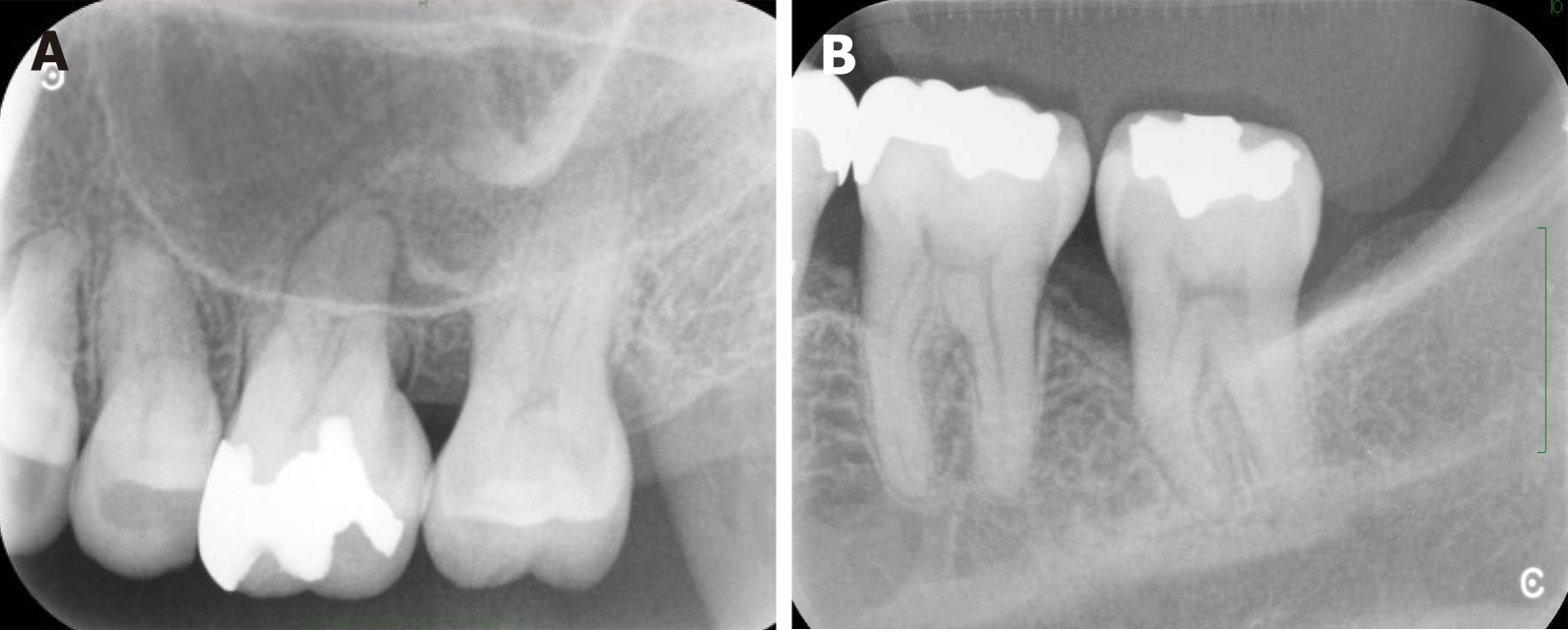
Figure 6 Dental radiographs of the upper and lower left molars after orthodontic treatment.
A: Upper left molars; B: Lower left molars.
Figure 7 Post-treatment facial photographs.
A: Lateral view; B: Front view.
Figure 8 Post-treatment intraoral photographs.
A: Right side view; B: Front view; C: Left side view; D: Incisal view; E: Upper occlusal view; F: Lower occlusal view.
Figure 9 Post-treatment panoramic radiograph.
Figure 10 Post-treatment cephalometric radiograph.
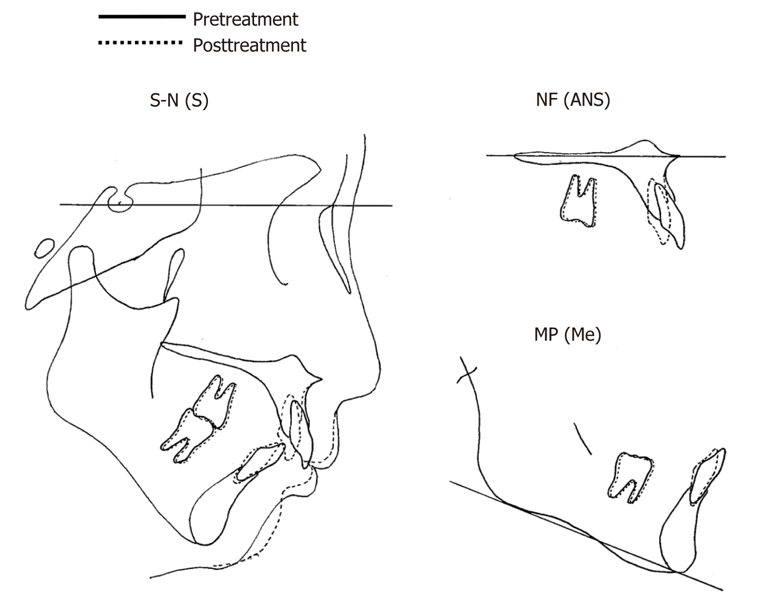
Figure 11 Cephalometric superimposition (pre-treatment and post-treatment).
S-N(S): Superimposition for sella-nasion at sella; NF(ANS): Superimposition for nasal floor at anterior nasal spine; MP(Me): Superimposition for mandibular plane at menton.
Figure 12 Dental radiographs of the upper and lower left molars after periodontal surgery.
A: Upper left molars; B: Lower left molars.
Figure 13 Intraoral photographs after 10 yr of retention.
A: Right side view; B: Front view; C: Left side view; D: Incisal view; E: Upper occlusal view; F: Lower occlusal view.
- Citation: Kaku M, Matsuda S, Kubo T, Shimoe S, Tsuga K, Kurihara H, Tanimoto K. Generalized periodontitis treated with periodontal, orthodontic, and prosthodontic therapy: A case report. World J Clin Cases 2021; 9(21): 6110-6124
- URL: https://www.wjgnet.com/2307-8960/full/v9/i21/6110.htm
- DOI: https://dx.doi.org/10.12998/wjcc.v9.i21.6110


