Copyright
©The Author(s) 2021.
World J Clin Cases. Jul 26, 2021; 9(21): 6081-6090
Published online Jul 26, 2021. doi: 10.12998/wjcc.v9.i21.6081
Published online Jul 26, 2021. doi: 10.12998/wjcc.v9.i21.6081
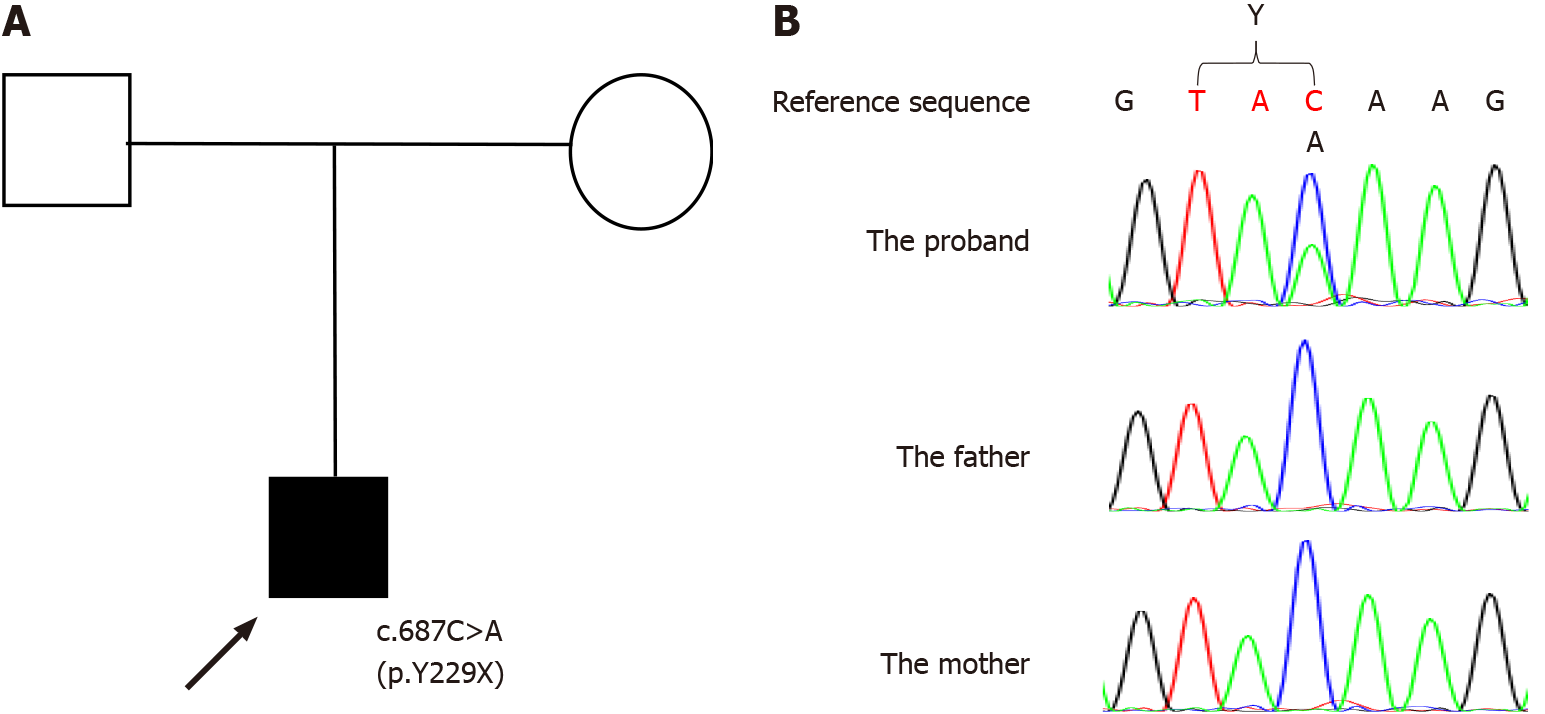
Figure 1 The family pedigree and mutations detected in SATB2.
A: The pedigree of the family with SATB2-associated syndrome (SAS). The arrow indicates the proband; the parents have no signs of SATB2-associated syndrome; B: The mutations detected in the family. The mutation is de novo in the proband, whereas the parents are wild-type.
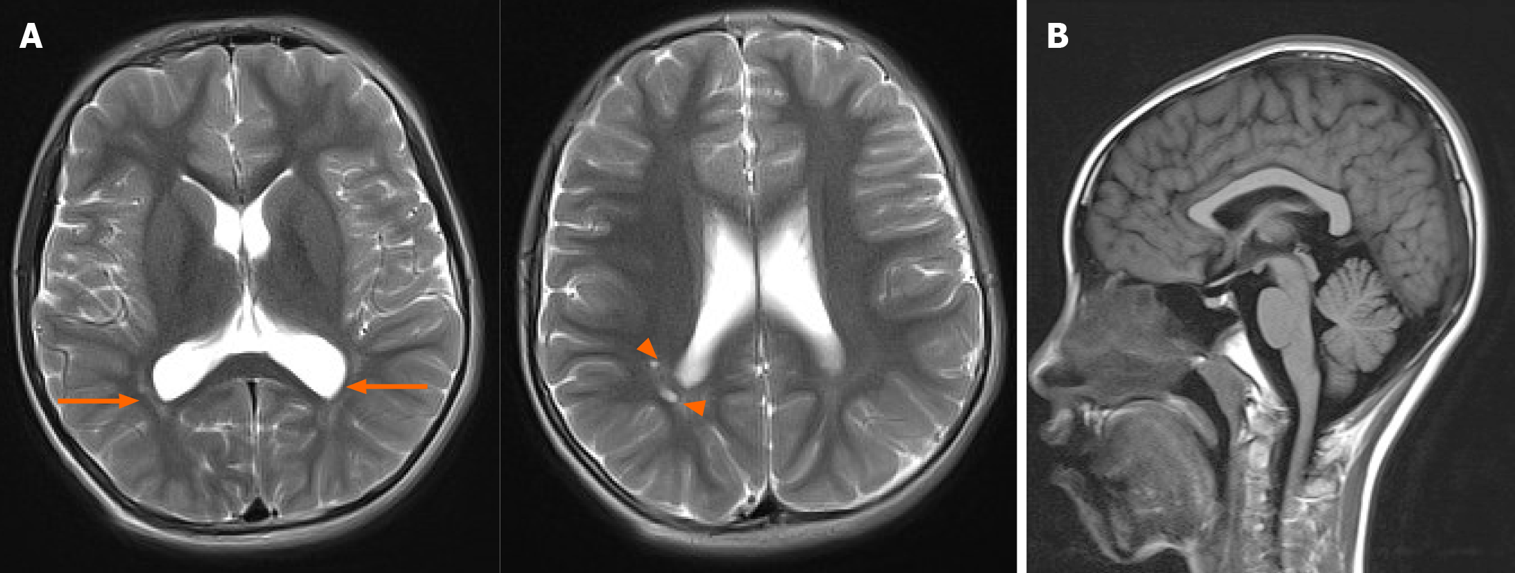
Figure 2 Brain magnetic resonance imaging images.
A: Multiple small cystic lesions in the white matter adjacent to the posterior horns of the right lateral ventricle (arrowheads) and long T2 signals in the white matter adjacent to the bilateral posterior horns of the lateral ventricles were found in transversal T2-weighted images (arrows); B: The corpus callosum was found normal in sagittal fluid-attenuated inversion recovery image.
- Citation: Zhu YY, Sun GL, Yang ZL. SATB2-associated syndrome caused by a novel SATB2 mutation in a Chinese boy: A case report and literature review. World J Clin Cases 2021; 9(21): 6081-6090
- URL: https://www.wjgnet.com/2307-8960/full/v9/i21/6081.htm
- DOI: https://dx.doi.org/10.12998/wjcc.v9.i21.6081


