Copyright
©The Author(s) 2021.
World J Clin Cases. Jul 26, 2021; 9(21): 6017-6025
Published online Jul 26, 2021. doi: 10.12998/wjcc.v9.i21.6017
Published online Jul 26, 2021. doi: 10.12998/wjcc.v9.i21.6017
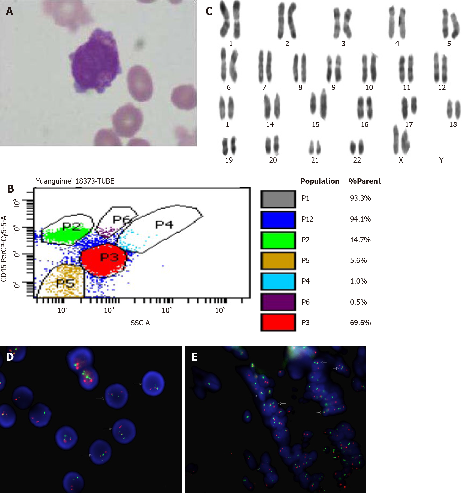
Figure 1 Laboratory examination results of this patient.
A: Bone marrow morphology revealed 68% of abnormal promyelocyte cells (100 ×); B: Flow cytometric analysis of bone marrow cells showed a population of abnormal cells (P3: 69.6%); C: Karyotype of this patient: 46, XX, t (15; 17) (q22; q21); D: Fluorescence in situ hybridization (FISH) results in bone marrow cells at diagnosis showed PML/RARα fusion; E: FISH results in intestinal tissue cells at diagnosis showed PML/RARα fusion.
- Citation: Wang L, Cai DL, Lin N. Myeloid sarcoma of the colon as initial presentation in acute promyelocytic leukemia: A case report and review of the literature. World J Clin Cases 2021; 9(21): 6017-6025
- URL: https://www.wjgnet.com/2307-8960/full/v9/i21/6017.htm
- DOI: https://dx.doi.org/10.12998/wjcc.v9.i21.6017


