Copyright
©The Author(s) 2021.
World J Clin Cases. Jul 26, 2021; 9(21): 5988-5998
Published online Jul 26, 2021. doi: 10.12998/wjcc.v9.i21.5988
Published online Jul 26, 2021. doi: 10.12998/wjcc.v9.i21.5988
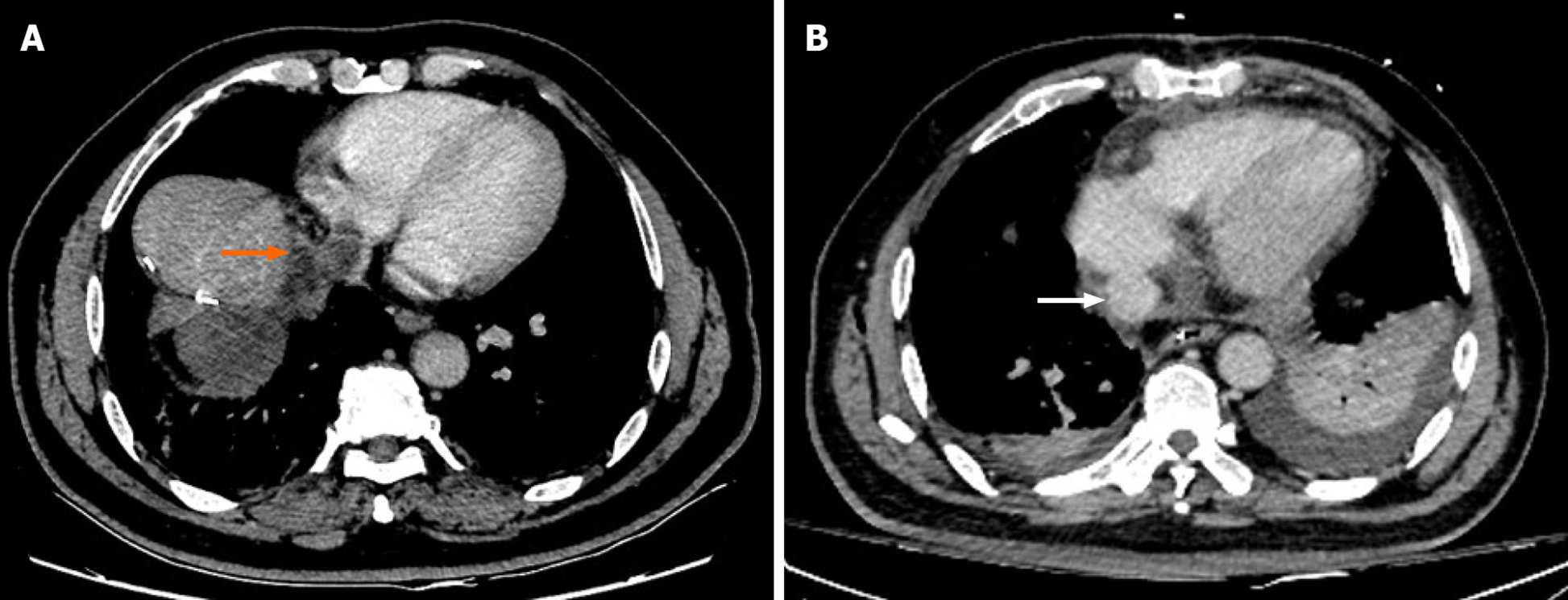
Figure 1 Computed tomography images of inferior vena cava tumor thrombus before and after a second surgery.
A: Acquired before surgery showed the existence of an inferior vena cava tumor thrombus that protruded into the right atrium (orange arrow); B: Acquired after surgery showed that the tumor thrombus was removed (white arrow).
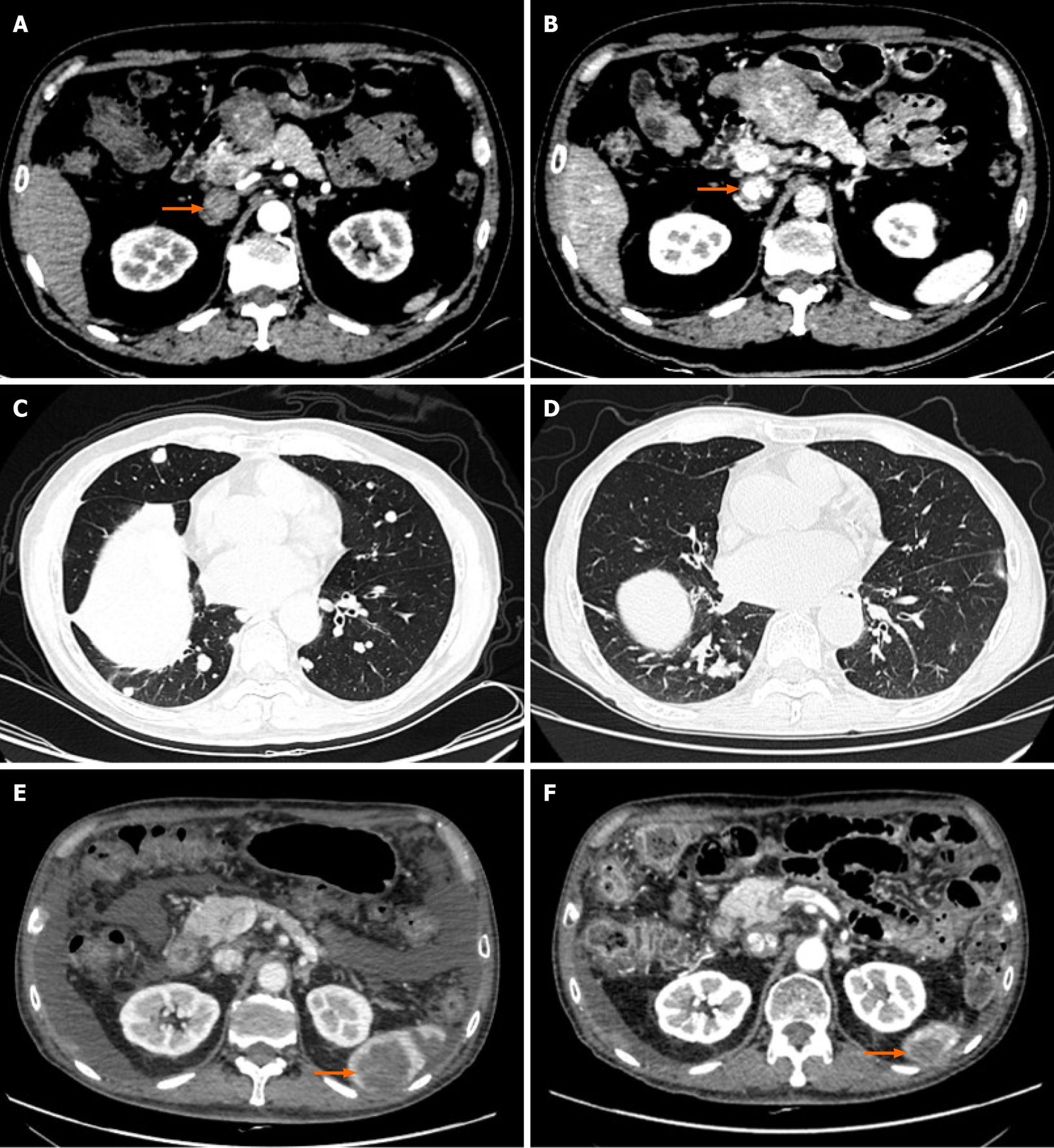
Figure 2 Computed tomography images of inferior vena cava tumor thrombus and lung and splenic metastases.
A and B: Acquired before surgery showed the existence of inferior vena cava tumor thrombus (orange arrows); C: Acquired before immunotherapy showed the existence of lung metastases; D: Acquired after immunotherapy showed that the lung metastases were stable; E: Acquired before immunotherapy showed the existence of a splenic metastasis (orange arrow); F: Acquired after immunotherapy showed that the splenic metastasis was smaller (orange arrow).
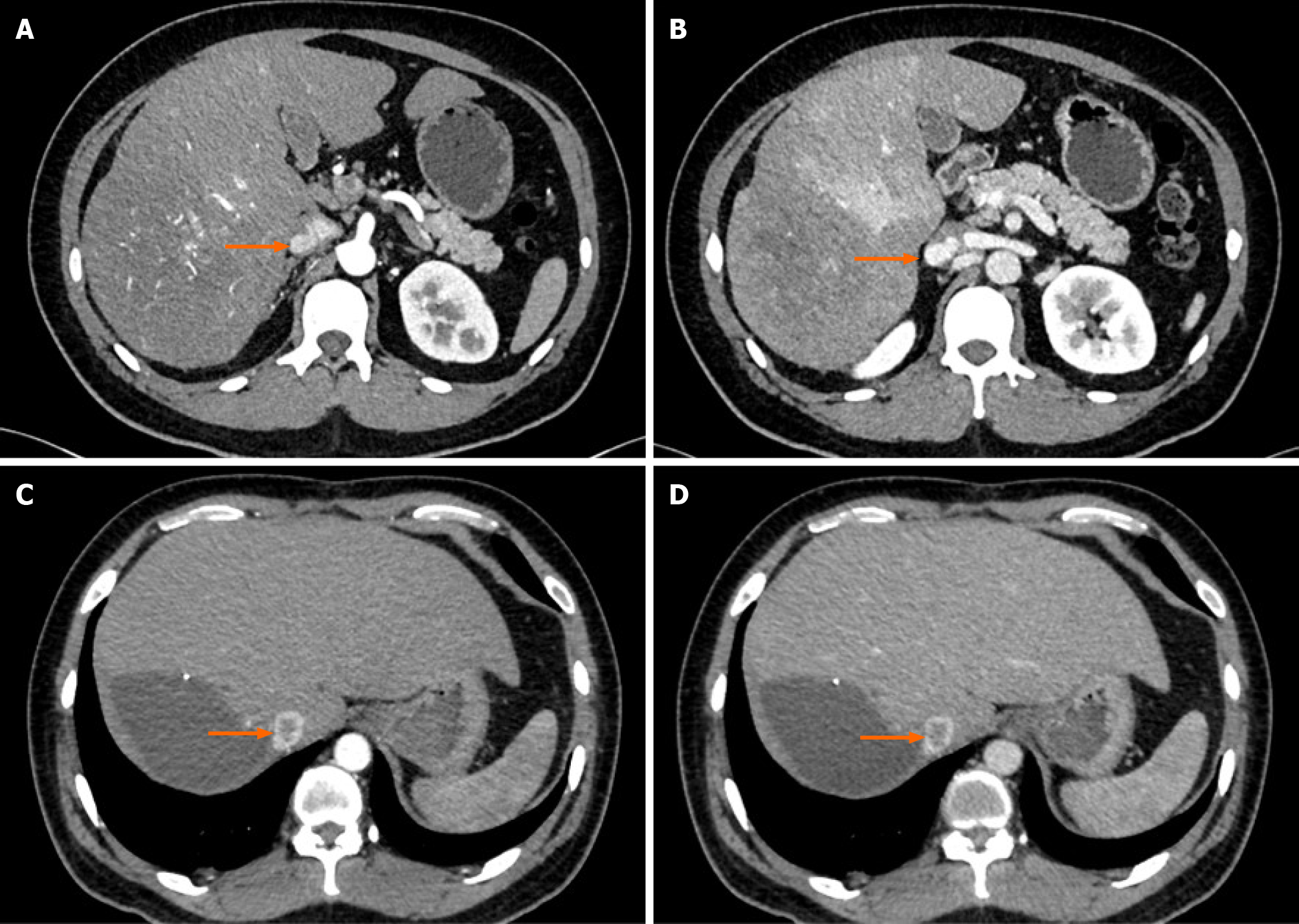
Figure 3 Computed tomography images of inferior vena cava tumor thrombus.
A and B: The inferior vena cava tumor thrombus (IVCTT) before surgery (orange arrows); C and D: The IVCTT after two courses of immunotherapy (orange arrows).
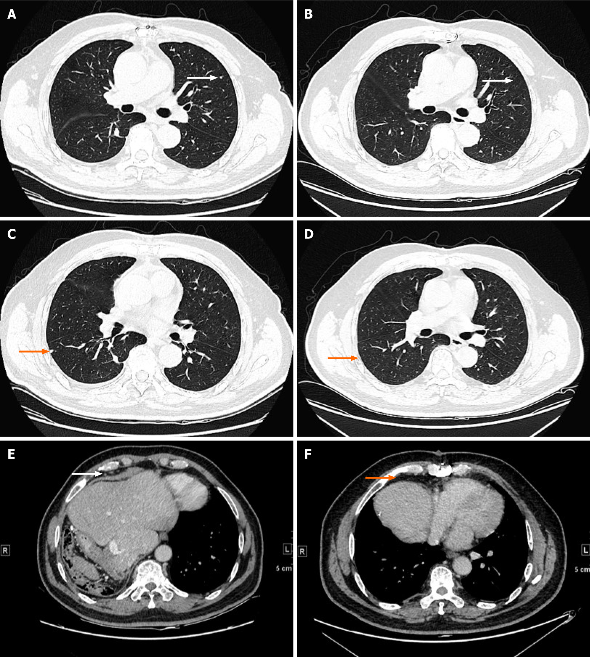
Figure 4 Computed tomography images of lung metastases and mediastinal lymph node metastases before and after immunotherapy.
A and C: Acquired before immunotherapy showed the existence of lung metastases (arrows); B and D: Acquired after immunotherapy showed that the lung metastases had disappeared (arrows); E: Acquired before immunotherapy showed the existence of mediastinal lymph node metastases (white arrow); F: Acquired after immunotherapy showed that the mediastinal lymph node metastases became smaller (orange arrow).
- Citation: Liu SR, Yan Q, Lin HM, Shi GZ, Cao Y, Zeng H, Liu C, Zhang R. Anti-programmed cell death ligand 1-based immunotherapy in recurrent hepatocellular carcinoma with inferior vena cava tumor thrombus and metastasis: Three case reports. World J Clin Cases 2021; 9(21): 5988-5998
- URL: https://www.wjgnet.com/2307-8960/full/v9/i21/5988.htm
- DOI: https://dx.doi.org/10.12998/wjcc.v9.i21.5988


