Copyright
©The Author(s) 2021.
World J Clin Cases. Jul 26, 2021; 9(21): 5980-5987
Published online Jul 26, 2021. doi: 10.12998/wjcc.v9.i21.5980
Published online Jul 26, 2021. doi: 10.12998/wjcc.v9.i21.5980
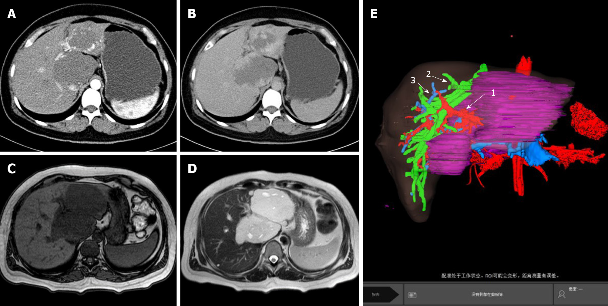
Figure 1 Image data before operative treatments.
A and B: Computed tomography images before operative treatment; C and D: Magnetic resonance imaging images before operative treatment; E: Liver volume model (E1: Hepatic artery; E2: Hepatic vein; E3: Portal vein).
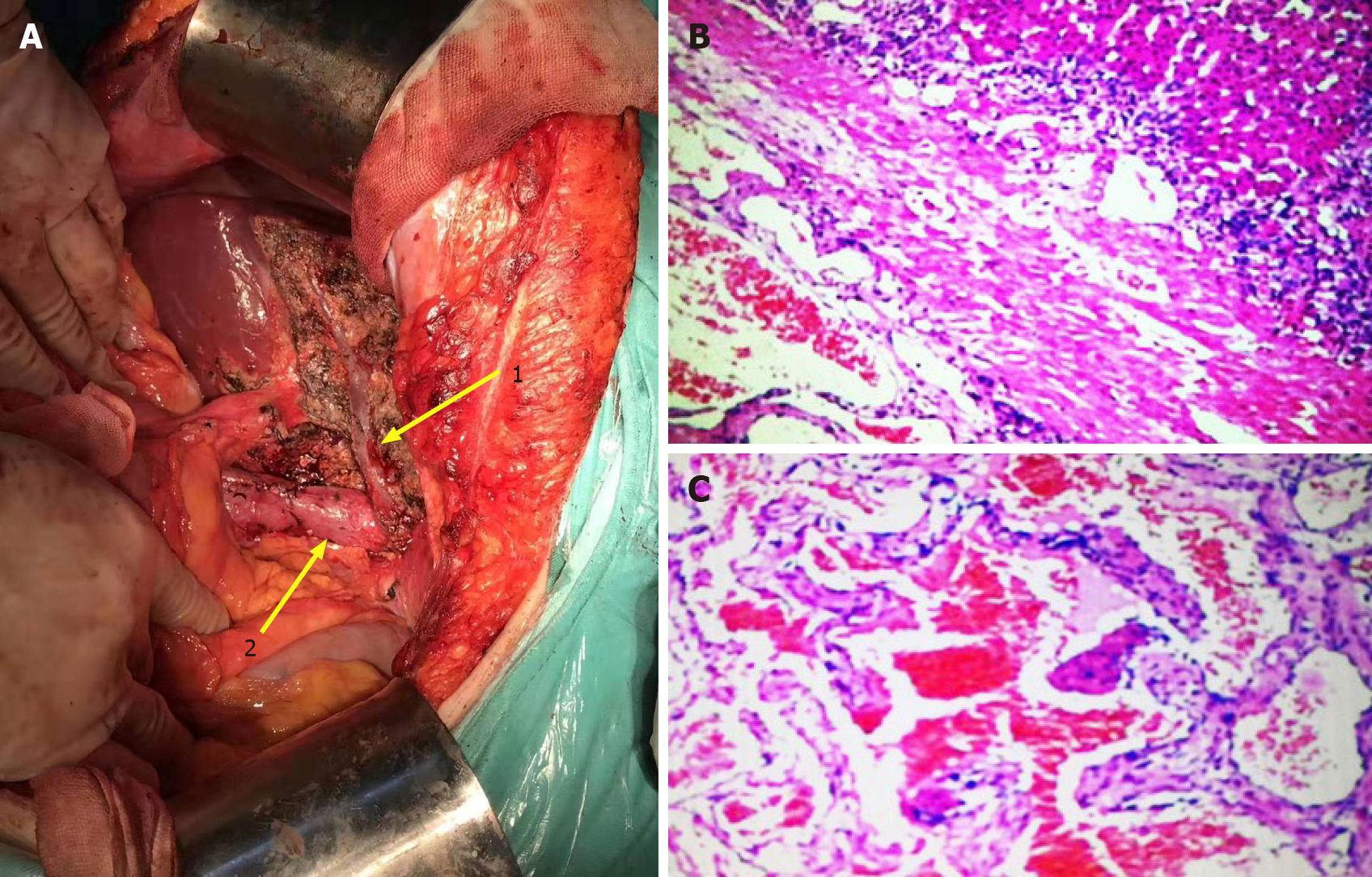
Figure 2 Images from the operation and pathology images after operative treatment.
A1: Middle hepatic vein; A2: Inferior vena cava; B and C: Cavernous hemangioma.
- Citation: Wang XX, Dong BL, Wu B, Chen SY, He Y, Yang XJ. Giant hemangioma of the caudate lobe of the liver with surgical treatment: A case report. World J Clin Cases 2021; 9(21): 5980-5987
- URL: https://www.wjgnet.com/2307-8960/full/v9/i21/5980.htm
- DOI: https://dx.doi.org/10.12998/wjcc.v9.i21.5980


