Copyright
©The Author(s) 2021.
World J Clin Cases. Jul 26, 2021; 9(21): 5972-5979
Published online Jul 26, 2021. doi: 10.12998/wjcc.v9.i21.5972
Published online Jul 26, 2021. doi: 10.12998/wjcc.v9.i21.5972
Figure 1 Computed tomography of the chest demonstrated large right pleural effusion.
A: Lung window; B: Mediastinal window.
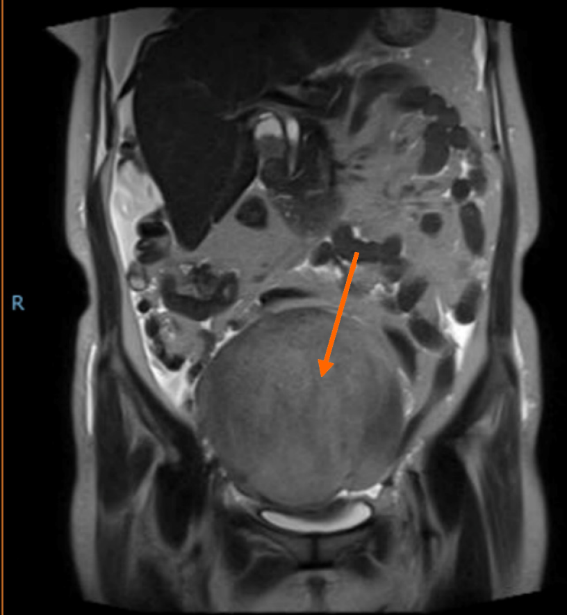
Figure 2
Magnetic resonance imaging showing a large ovarian tumor (arrows) on the right side of the pelvis.
Figure 3 Surgical pathology demonstrating a theca cell tumor of the right ovary (Hematoxylin and eosin staining).
A: 40 × magnification; B: 100 × magnification.
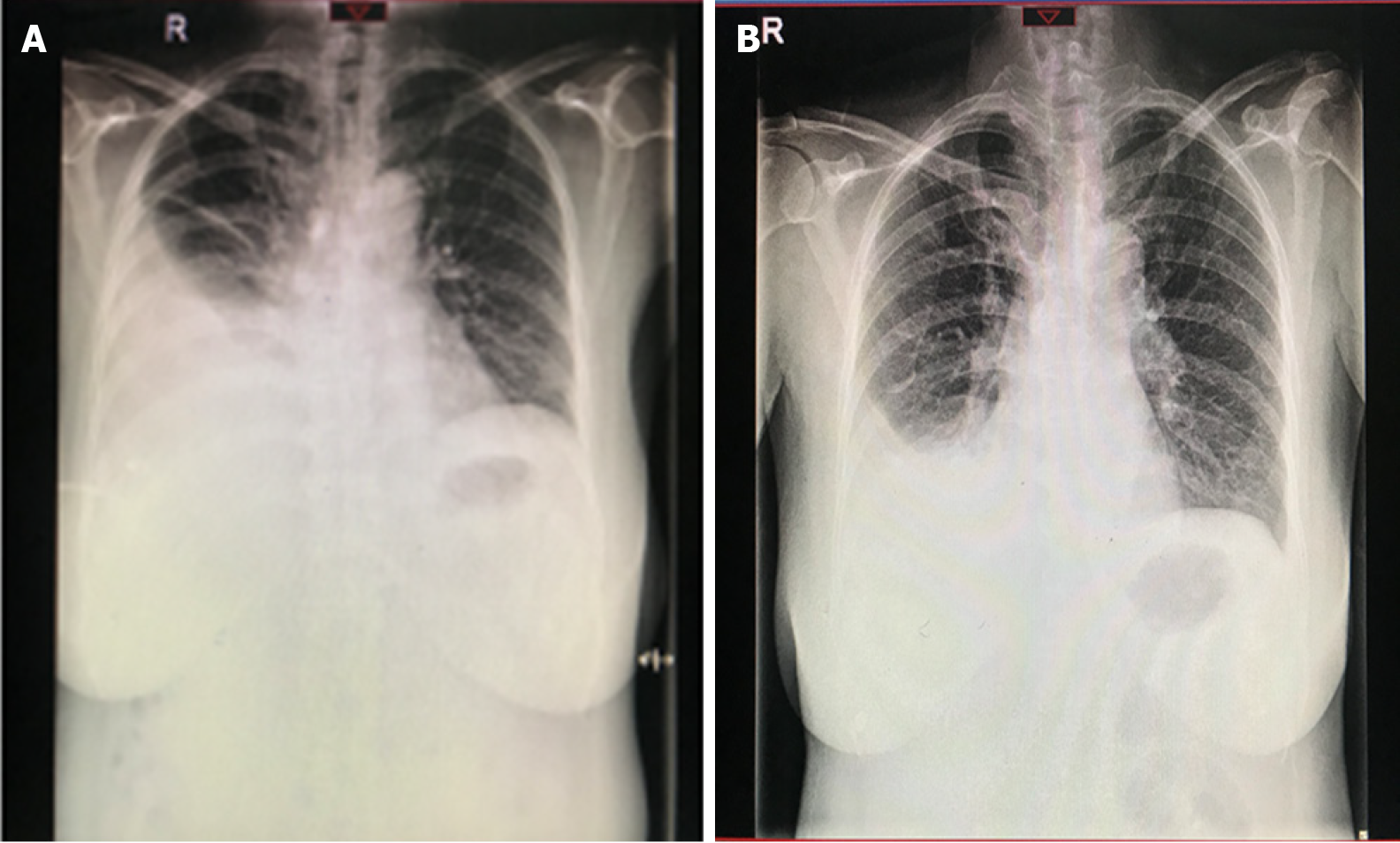
Figure 4 X-ray photographs demonstrating blunting of the right costophrenic angle before surgery and 1 wk after surgery.
A: Before surgery; B: 1 wk after surgery.
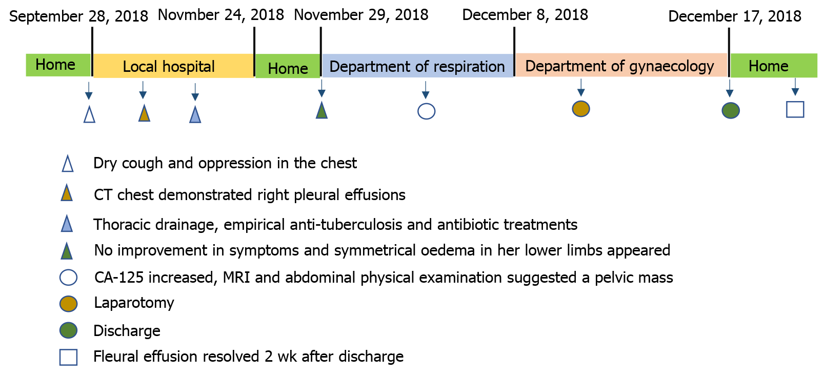
Figure 5 Therapy and course timeline of the patient.
MRI: Magnetic resonance imaging; CT: Computed tomography.
- Citation: Hou YY, Peng L, Zhou M. Meigs syndrome with pleural effusion as initial manifestation: A case report. World J Clin Cases 2021; 9(21): 5972-5979
- URL: https://www.wjgnet.com/2307-8960/full/v9/i21/5972.htm
- DOI: https://dx.doi.org/10.12998/wjcc.v9.i21.5972


