Copyright
©The Author(s) 2021.
World J Clin Cases. Jul 26, 2021; 9(21): 5963-5971
Published online Jul 26, 2021. doi: 10.12998/wjcc.v9.i21.5963
Published online Jul 26, 2021. doi: 10.12998/wjcc.v9.i21.5963
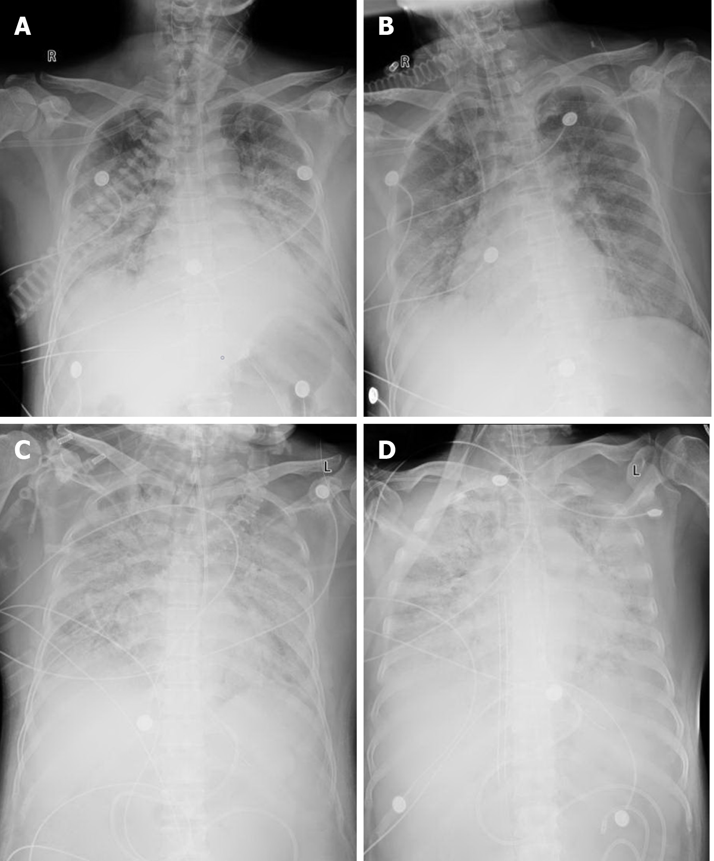
Figure 1 Chest X-ray images of a 53-year-old man with coronavirus disease 2019 at four time points.
A: Chest X-ray at admission; B: Chest X-ray after tracheal intubation; C: Chest X-ray after 96 h of mechanical ventilation; D: Chest X-ray after establishing extracorporeal membrane oxygenation.
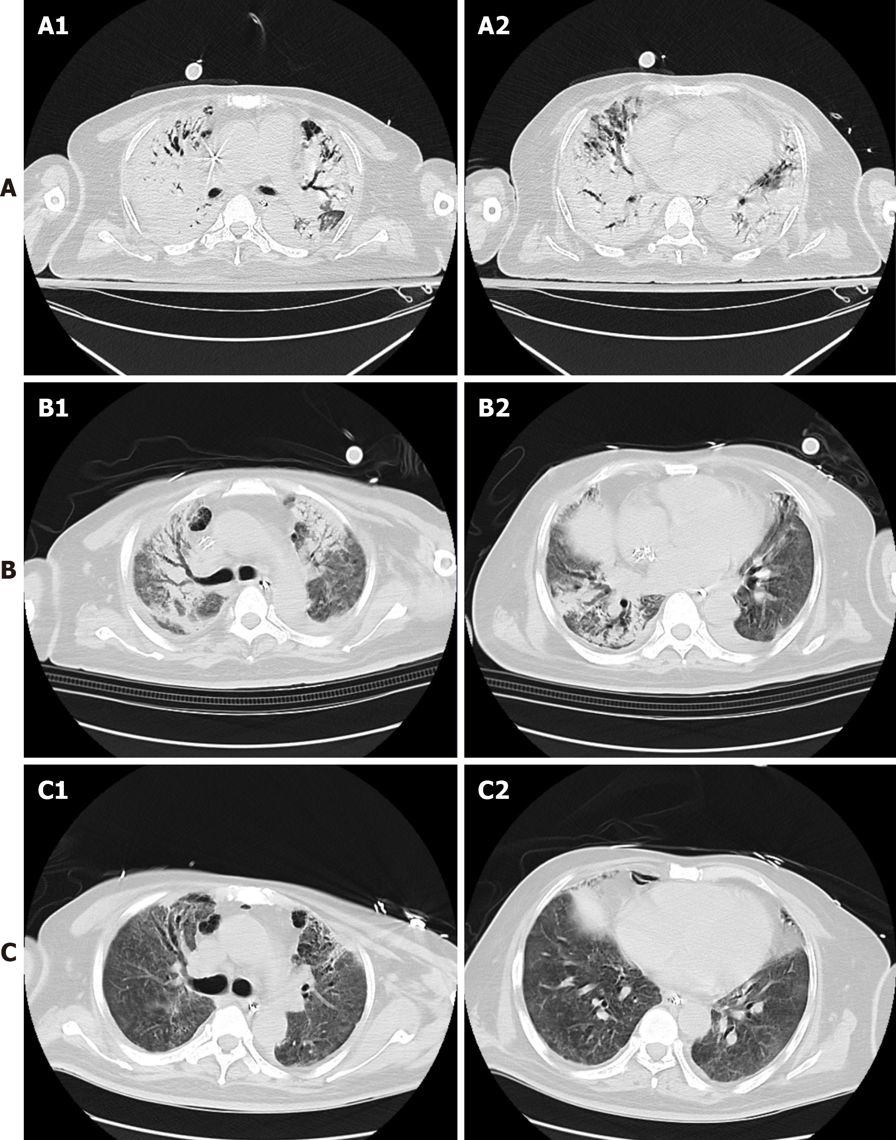
Figure 2 Chest computed tomography at three time points.
Two representative slices of the middle and lower lobe were chosen. A: 48 h of extracorporeal membrane oxygenation; B: 1 wk of extracorporeal membrane oxygenation and prone position ventilation; C: 5 d of awake extracorporeal membrane oxygenation and rehabilitation.
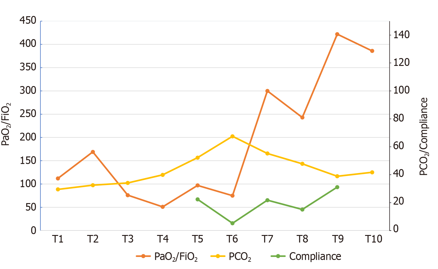
Figure 3 Changes in pressure of oxygen/fraction of inspiration O2 and pressure of carbon dioxide/compliance over time.
T1: Admission; T2: After 24 h of non-invasive ventilation; T3: Intensive care unit admission; T4: Before intubation; T5: After 24 h of intubation; T6: 24 h before extracorporeal membrane oxygenation (ECMO) was established; T7: Before ECMO weaning (ECMO oxygen concentration was 21%); T8: 24 h after ECMO weaning; T9: Before MV weaning; T10: 24 h after MV weaning. PaO2/FiO2: Pressure of oxygen/fraction of inspiration O2; PCO2: Pressure of carbon dioxide; MV: Mechanical ventilation.
- Citation: Zhang JC, Li T. Awake extracorporeal membrane oxygenation support for a critically ill COVID-19 patient: A case report. World J Clin Cases 2021; 9(21): 5963-5971
- URL: https://www.wjgnet.com/2307-8960/full/v9/i21/5963.htm
- DOI: https://dx.doi.org/10.12998/wjcc.v9.i21.5963


