Copyright
©The Author(s) 2021.
World J Clin Cases. Jul 16, 2021; 9(20): 5744-5751
Published online Jul 16, 2021. doi: 10.12998/wjcc.v9.i20.5744
Published online Jul 16, 2021. doi: 10.12998/wjcc.v9.i20.5744
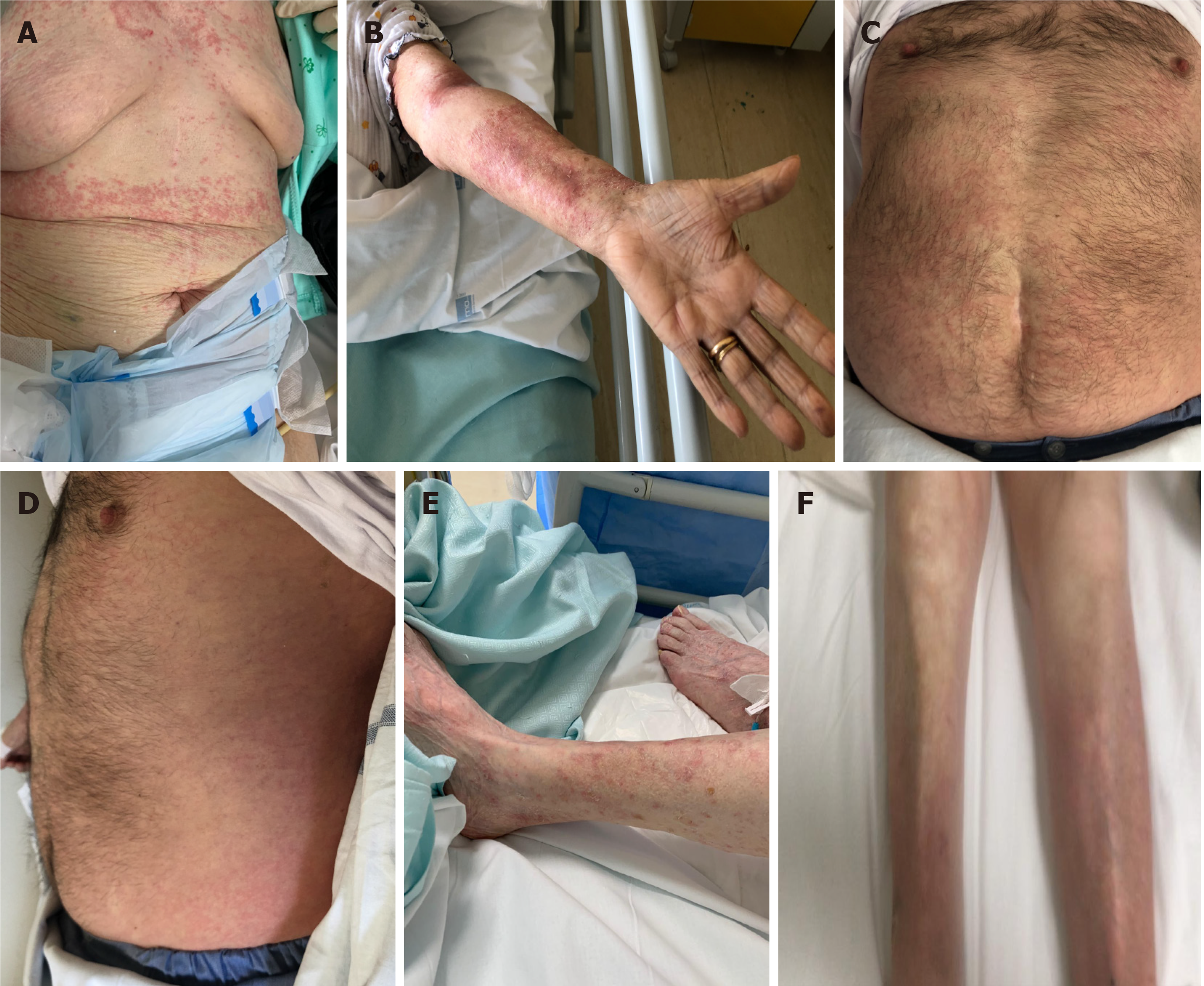
Figure 1 Clinical manifestations in coronavirus disease 2019 patients.
A and B: Thirty-eight days after the first positive swab, an erythematous and itchy skin rash appeared in patient 1 the sub-mammary region, rapidly extending to the trunk and the upper limbs; C and D: Twenty-eight days after the first positive swab for severe acute respiratory syndrome coronavirus 2 (SARS-CoV-2), an itchy and urticarial rash appeared in patient 2 on the chest and arms; E and F: Thirty-eight days after the first positive swab for SARS-CoV-2, patient 3 developed an itchy and erythematous rash on both the lower limbs.
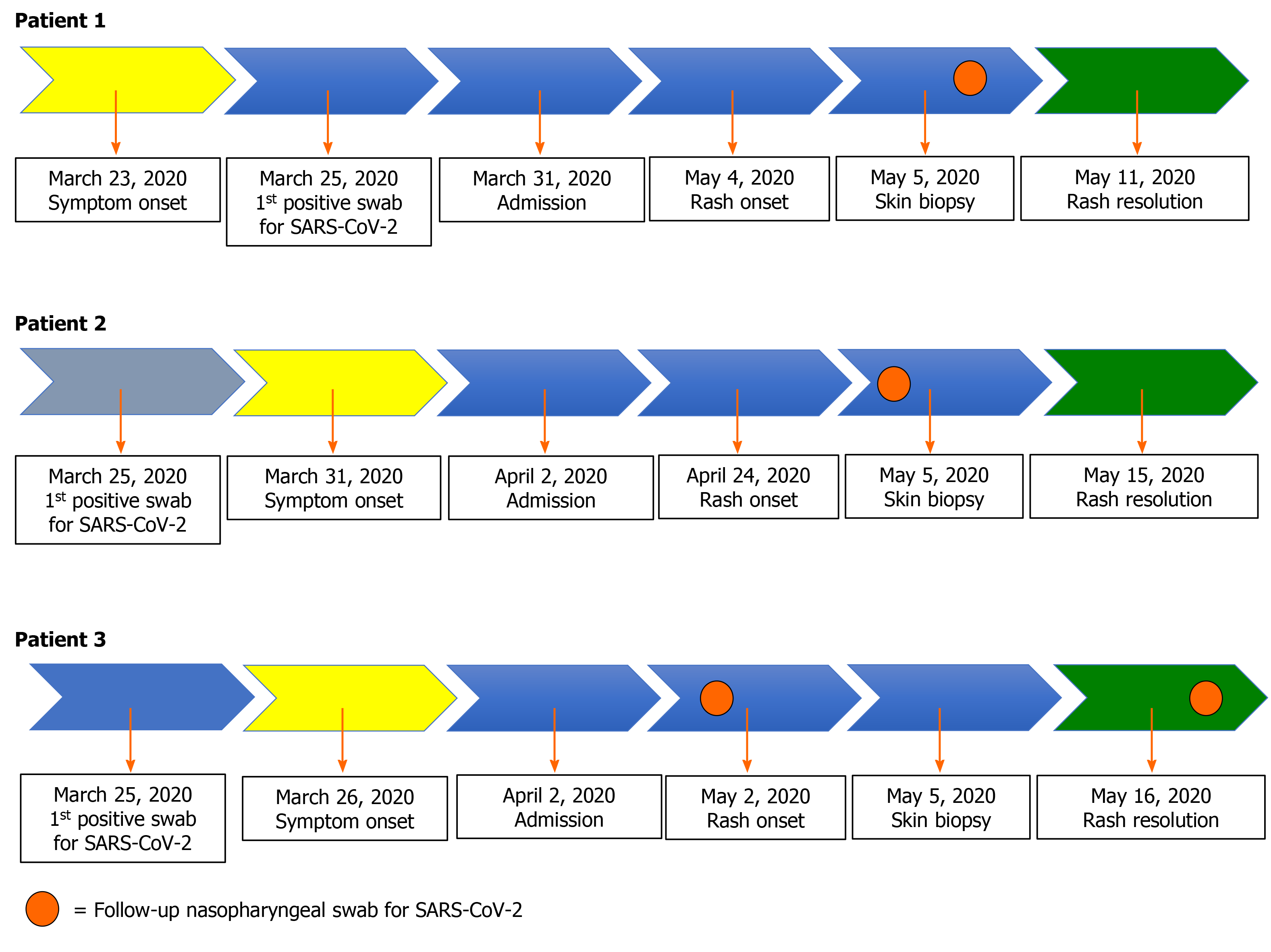
Figure 2 Clinical course for each patient.
For patient 1 the polymerase chain reaction (PCR) for severe acute respiratory syndrome coronavirus 2 (SARS-CoV-2) on nasopharyngeal swab performed on May 5 came back as positive for the N gene (33 cycle threshold). Similarly, for patient 2, the nasopharyngeal swab performed 2 d before the rash onset (April 22, 2020) also came back as positive for the N gene (36 cycle threshold). For patient 3, PCR for SARS-CoV-2 on nasopharyngeal swab performed a week before the rash onset was negative, while the one performed a week later (May 16, 2020, the same day of rash healing) was positive only for the N gene (37 cycle threshold).
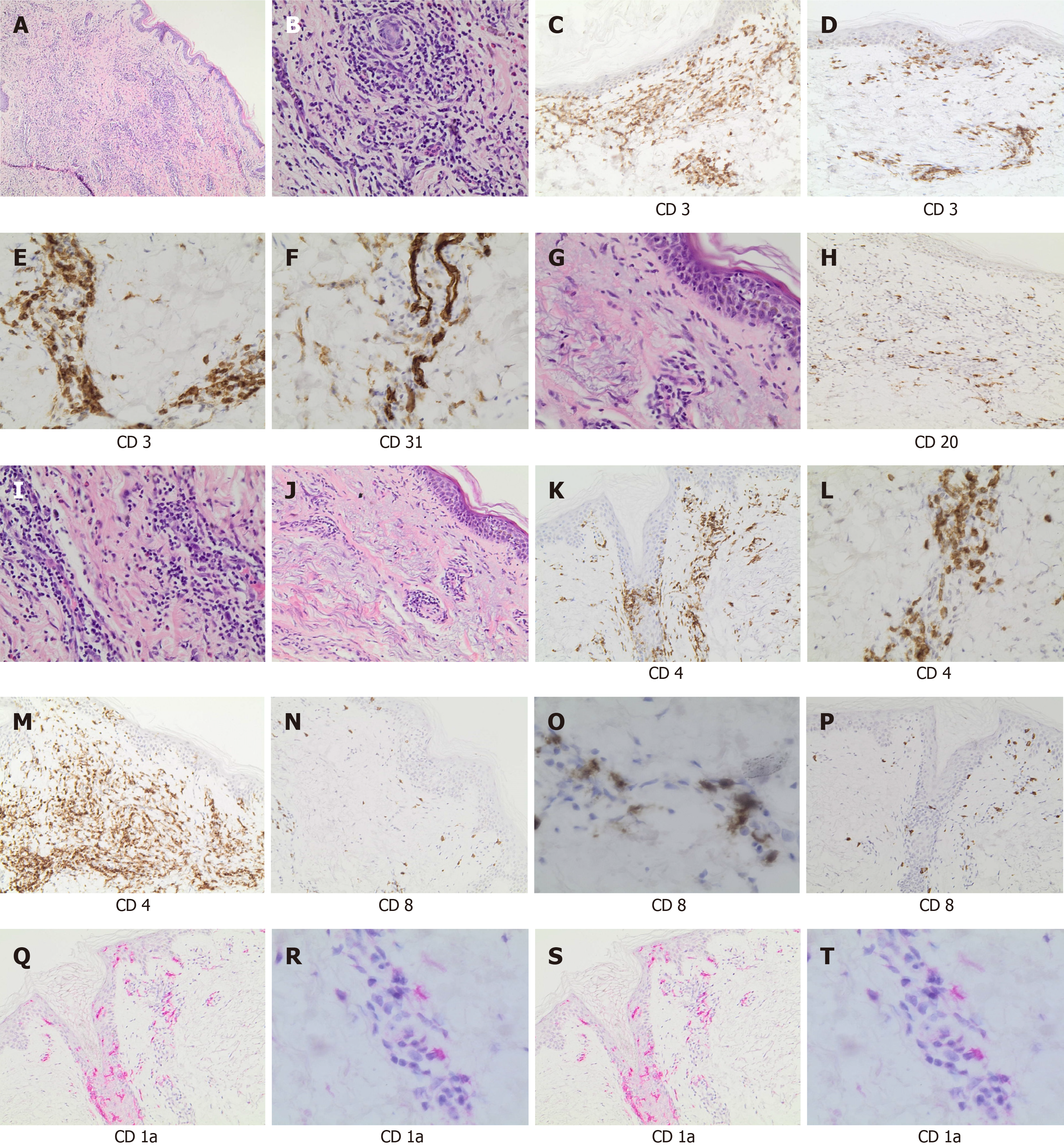
Figure 3 Histological section and immunophenotyping.
A: Panoramic view of cutaneous lesion showed chronic dermatitis with perivascular lymphocytic infiltrate (hematoxylin and eosin staining, × 40 magnification); B: A detail of perivascular infiltrate (× 200 magnification); C: CD3 staining highlighted the diffuse T-cell infiltrate (× 200 magnification); D: Rare B cell lymphocytes (CD20+) were present (× 200 magnification); E: CD4 staining showed the prevalence of the T-helper lymphocytes (× 200 magnification); F: Only rare CD8 positive lymphocytes were observed (× 200 magnification); G: Epidermotropism (single-cell exocytosis of lymphocytes into the epidermis) (hematoxylin and eosin staining, × 200 magnification); H: CD3 staining highlighted the presence of T cell infiltrate, perivascular and intra-epidermal (× 100 magnification); I: CD4 staining was prevalent and demonstrated the presence of T-helper lymphoid cells in the epidermidis (× 100 magnification); L: CD8 staining evidentiated only rare cytotoxic (× 100 magnification); M: Morphologic evaluation demonstrated vascular proliferation associated to vasculitic phenomena (hematoxylin and eosin staining, × 200 magnification); N: CD31 staining highlighted vascular component (× 200 magnification); O: CD3 staining showed T cell vascular/perivascular infiltrate (× 200 magnification); P: CD4 staining indicated the prevalence of T-helper lymphoid cells in perivascular and vascular component (× 200 magnification); Q: CD8 staining indicated only rare cytotoxic (× 100 magnification); R: Morphologic evaluation showed a chronic dermatitis with perivascular infiltrate (hematoxylin and eosin staining, × 40 magnification); S: CD1a staining highlighted Langerhans cell intra-epidermal component (× 100 magnification); T: CD1a staining showed the presence of Langerhans cell in vascular/perivascular infiltrate (× 200 magnification).
- Citation: Mazzitelli M, Dastoli S, Mignogna C, Bennardo L, Lio E, Pelle MC, Trecarichi EM, Pereira BI, Nisticò SP, Torti C. Histopathology and immunophenotyping of late onset cutaneous manifestations of COVID-19 in elderly patients: Three case reports. World J Clin Cases 2021; 9(20): 5744-5751
- URL: https://www.wjgnet.com/2307-8960/full/v9/i20/5744.htm
- DOI: https://dx.doi.org/10.12998/wjcc.v9.i20.5744


