Copyright
©The Author(s) 2021.
World J Clin Cases. Jul 16, 2021; 9(20): 5717-5723
Published online Jul 16, 2021. doi: 10.12998/wjcc.v9.i20.5717
Published online Jul 16, 2021. doi: 10.12998/wjcc.v9.i20.5717
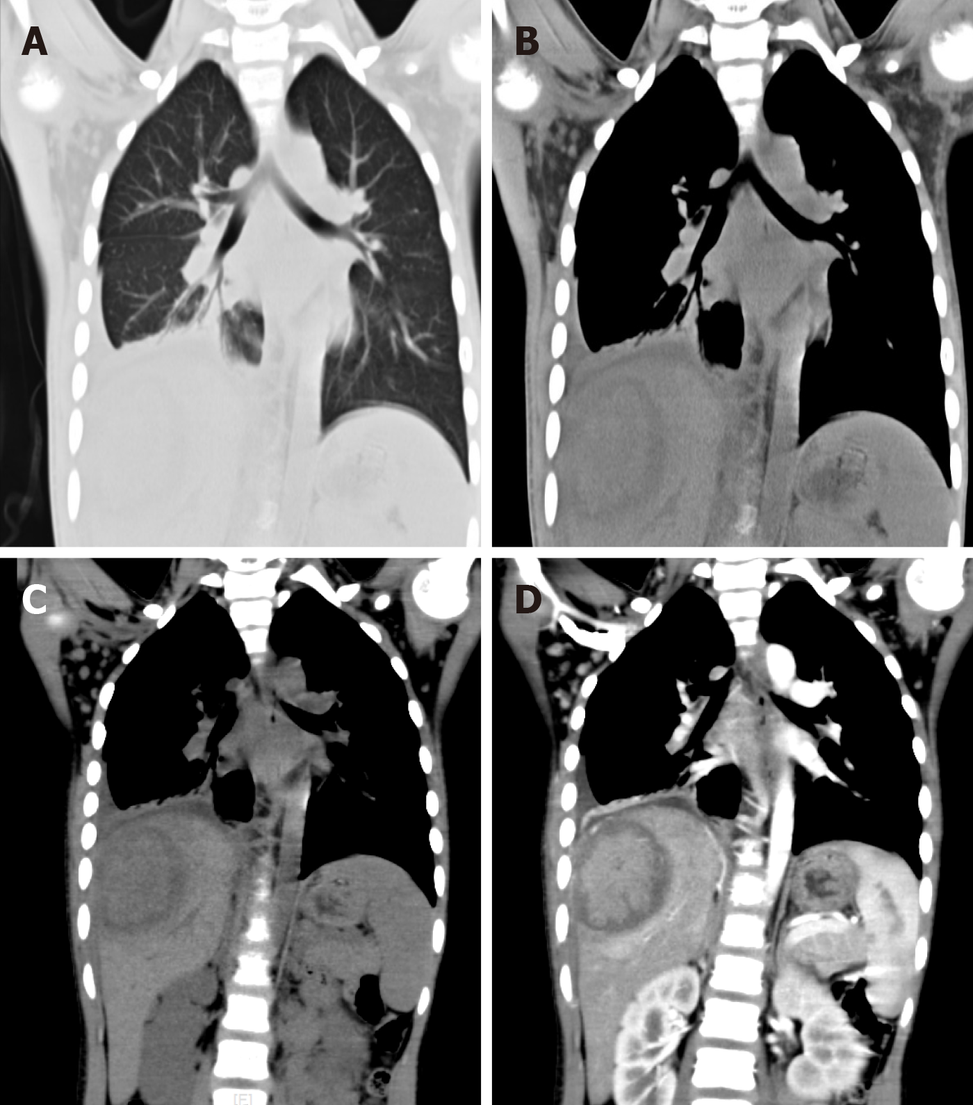
Figure 1 Chest and abdominal computed tomography images before antibiotic treatment.
A and B: The lung window (A) and soft-tissue (B) window of chest computed tomography (CT). Patchy enhanced density with blurred edges and uneven density showed in the right lower lung, with pleural effusion and pleural thickening on the right side. Soft tissue swelling was displayed in the right thoracic and abdominal wall; C and D: Plain scan (C) and arterial phase (D) of abdominal CT. The lesion was 5.2 cm × 7.2 cm × 5.5 cm in the S7 segment of the liver. The central density of the lesion was slightly lower, but the central enhancement of the lesion on arterial phase was obvious surrounded by a low-density ring. Hyperperfusion is seen around the lesion.
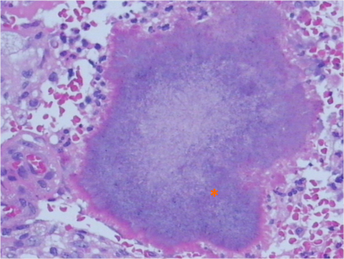
Figure 2 Histological diagnosis showing Actinomyces colonies (asterisk) in sample tissue (hematoxylin and eosin stain, original magnification × 400).
- Citation: Liang ZJ, Liang JK, Chen YP, Chen Z, Wang Y. Primary liver actinomycosis in a pediatric patient: A case report and literature review. World J Clin Cases 2021; 9(20): 5717-5723
- URL: https://www.wjgnet.com/2307-8960/full/v9/i20/5717.htm
- DOI: https://dx.doi.org/10.12998/wjcc.v9.i20.5717


