Copyright
©The Author(s) 2021.
World J Clin Cases. Jul 16, 2021; 9(20): 5695-5700
Published online Jul 16, 2021. doi: 10.12998/wjcc.v9.i20.5695
Published online Jul 16, 2021. doi: 10.12998/wjcc.v9.i20.5695
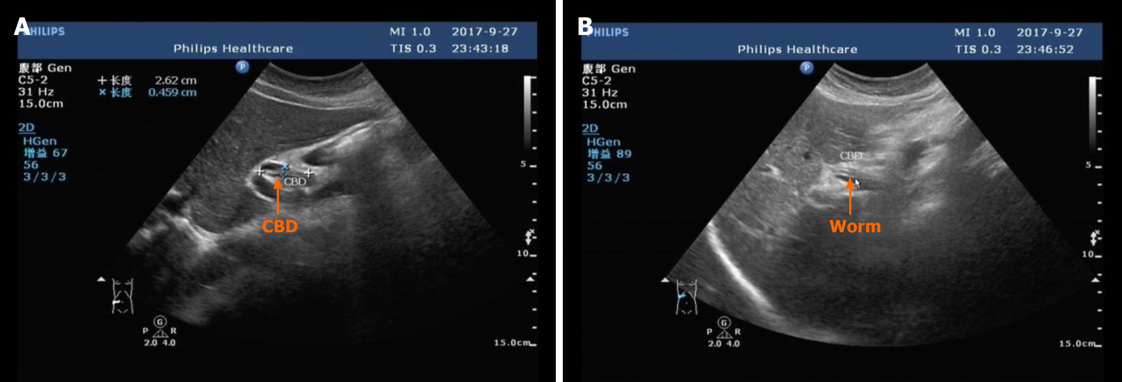
Figure 1 Abdominal ultrasonic diagnosis.
A: Abdominal ultrasound showed a slight dilation of upper part of the common bile duct; B: Abdominal ultrasound showed parallel tube-like structures (orange arrow). The common bile duct is dilated, about 8 mm in diameter.
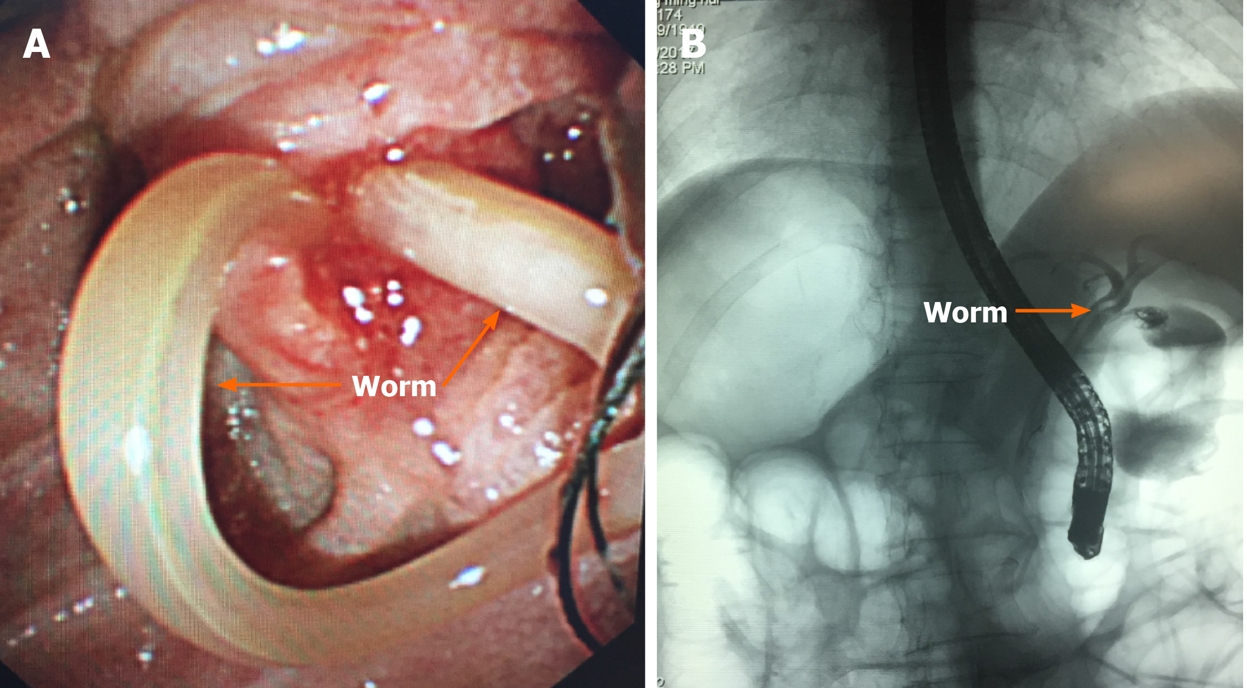
Figure 2 Endoscopic retrograde cholangiopancreatography showed that the intrahepatic bile duct was not dilated, and an active worm shadow was seen from the common bile duct to the right hepatic duct.
A: Common bile duct (orange arrow); B: Right hepatic duct (orange arrow).
- Citation: Wang X, Lv YL, Cui SN, Zhu CH, Li Y, Pan YZ. Endoscopic management of biliary ascariasis: A case report. World J Clin Cases 2021; 9(20): 5695-5700
- URL: https://www.wjgnet.com/2307-8960/full/v9/i20/5695.htm
- DOI: https://dx.doi.org/10.12998/wjcc.v9.i20.5695


