Copyright
©The Author(s) 2021.
World J Clin Cases. Jul 16, 2021; 9(20): 5675-5682
Published online Jul 16, 2021. doi: 10.12998/wjcc.v9.i20.5675
Published online Jul 16, 2021. doi: 10.12998/wjcc.v9.i20.5675
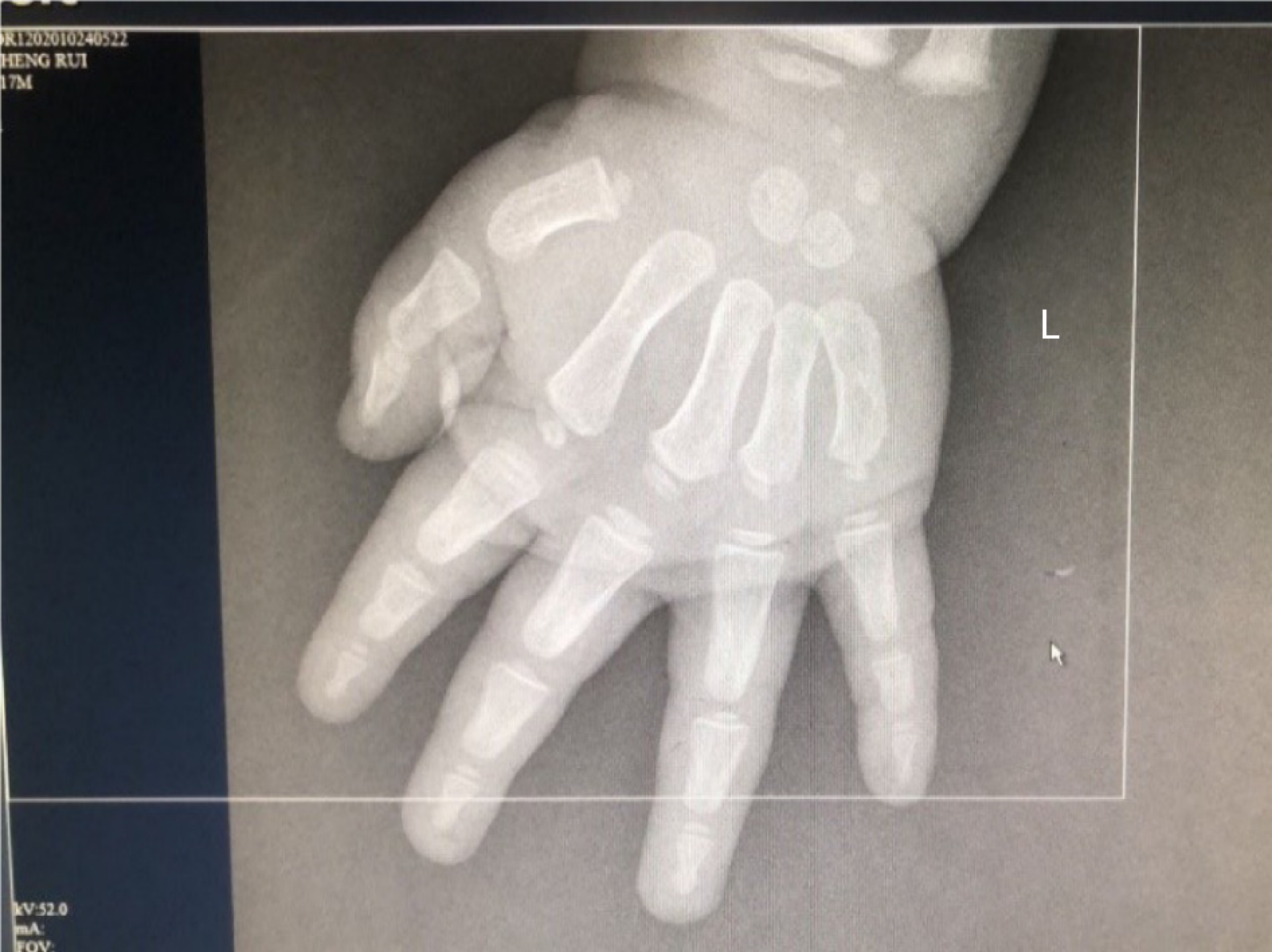
Figure 1 The ossification center indicates 3-year-old bone age changes.
Figure 2 Complete tumor capsule, tumor size 5.
5 cm × 5.0 cm × 3.0 cm, no definite capsular invasion and no definite vein invasion were found.
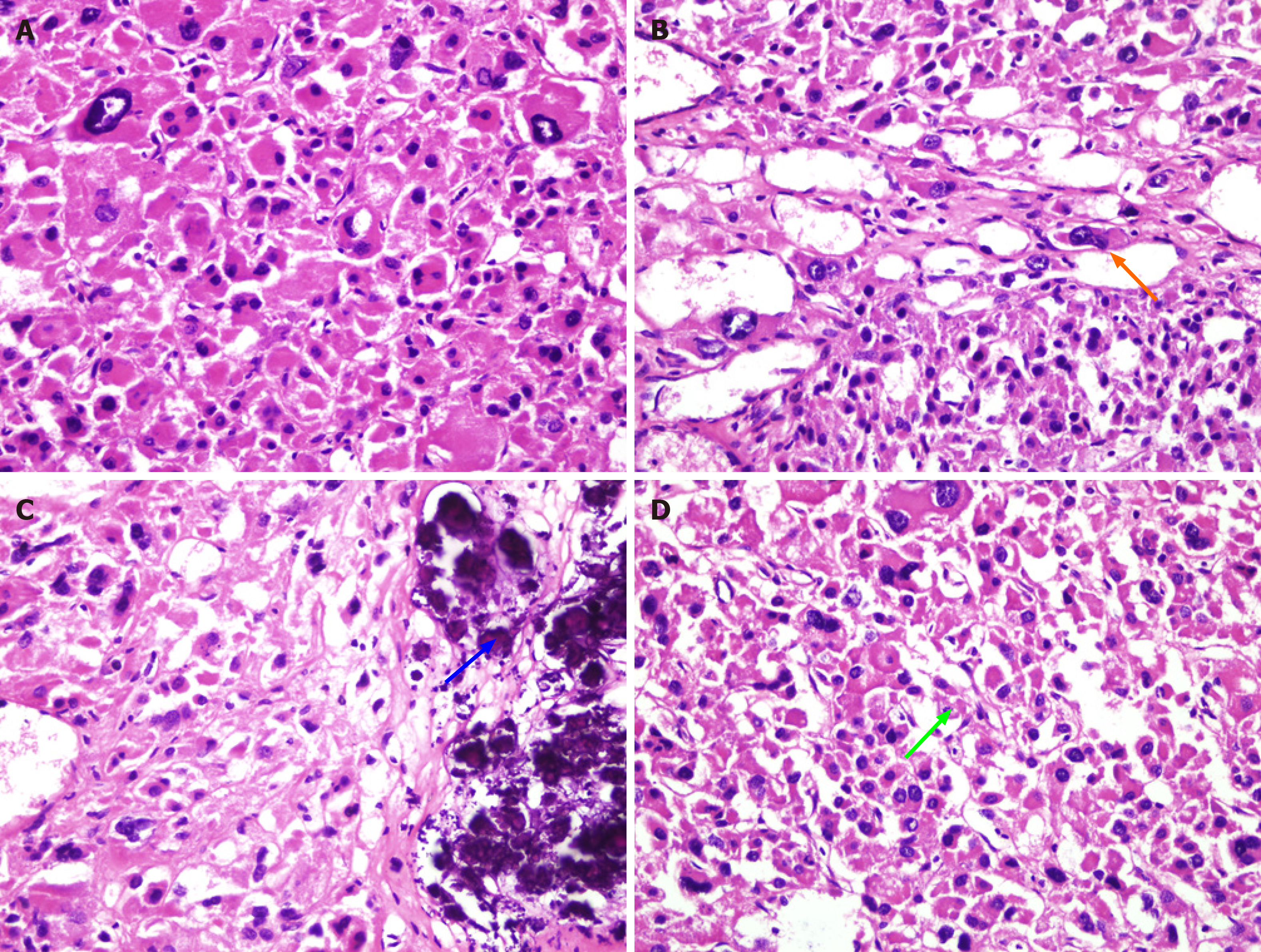
Figure 3 Eosinophils were predominant; visible wide cell atypical; no definite necrosis was found; no definite capsular invasion was found; no definite vein violation; visible focal sinus infiltration; some calcification is visible.
A: Typical eosinophilic changes (hematoxylin-eosin staining, image at 400 × magnification); B: This is a focal sinus infiltration of eosinophils (hematoxylin-eosin staining, image at 400 × magnification). Orange arrow: Focal sinus infiltration; C: Calcification: Blue fine particles aggregate (hematoxylin-eosin staining, image at 400 × magnification). Blue arrow: Calcification; D: Mitotic images count 1/10 hibernation-promoting factor (hematoxylin-eosin staining, image at 400 × magnification). Green arrow: Mitotic nuclear division.
- Citation: Chen XC, Tang YM, Mao Y, Qin DR. Oncocytic adrenocortical tumor with uncertain malignant potential in pediatric population: A case report and review of literature. World J Clin Cases 2021; 9(20): 5675-5682
- URL: https://www.wjgnet.com/2307-8960/full/v9/i20/5675.htm
- DOI: https://dx.doi.org/10.12998/wjcc.v9.i20.5675


