Copyright
©The Author(s) 2021.
World J Clin Cases. Jul 16, 2021; 9(20): 5668-5674
Published online Jul 16, 2021. doi: 10.12998/wjcc.v9.i20.5668
Published online Jul 16, 2021. doi: 10.12998/wjcc.v9.i20.5668
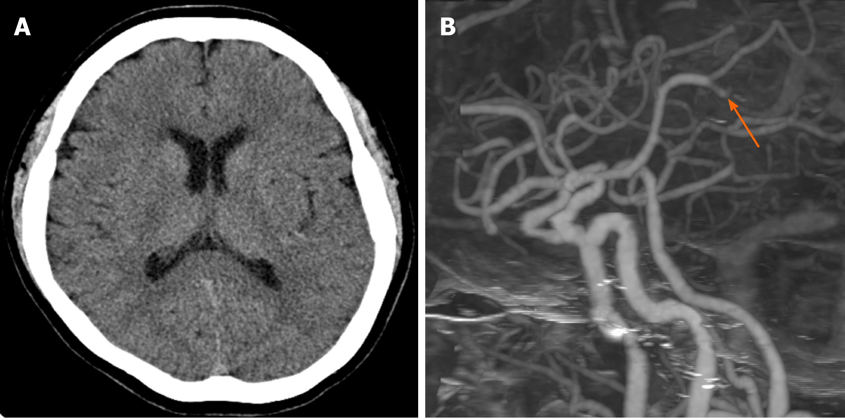
Figure 1 A 63-year-old woman presented with sudden onset of right hemiparesis and global aphasia (National Institutes of Health Stroke Scale = 15).
A: Initial brain computed tomography (CT) showed subtle decreased cortical densities at the left insula and temporal lobe, and CT ASPECT score was judged as 8 points; B: CT angiography showed occlusion of the inferior division of the M2-3 segment of the left middle cerebral artery (arrow).
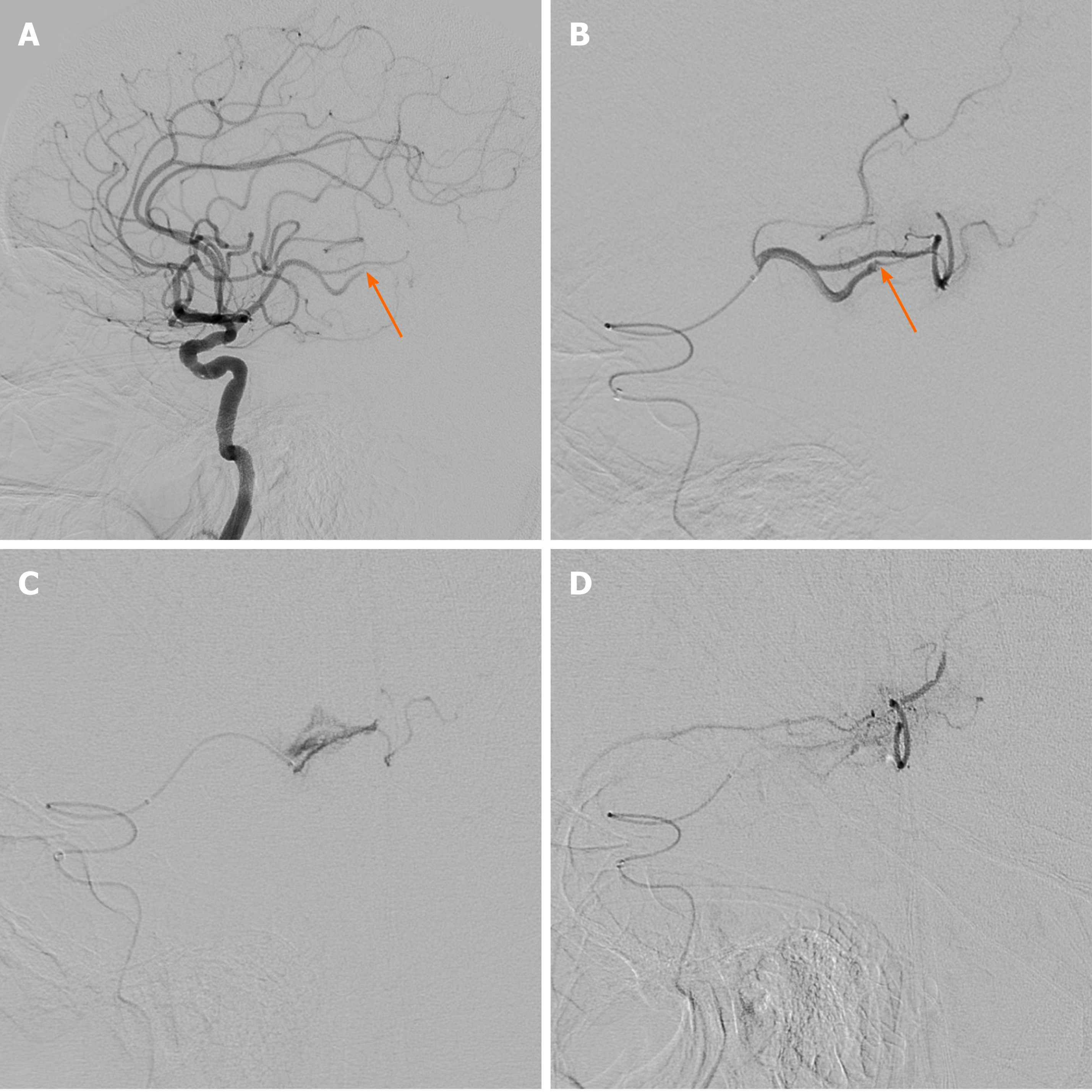
Figure 2 Cerebral angiography and endovascular management.
A and B: Initial cerebral angiography revealed occlusion of the inferior division of the middle cerebral artery at the M2-3 segment (arrow); C: During advancement of the microcatheter, resistance was encountered at the occlusion point, and the tension of the microcatheter was released. Cautious angiography was performed, revealing extravasation at the M3 branch due to vascular injury; D: Extravasation of contrast medium persisted in delayed angiography, and embolization was performed using gelfoam (1400-2000 μm). Repeat angiography confirmed hemostasis of the injured vessel.
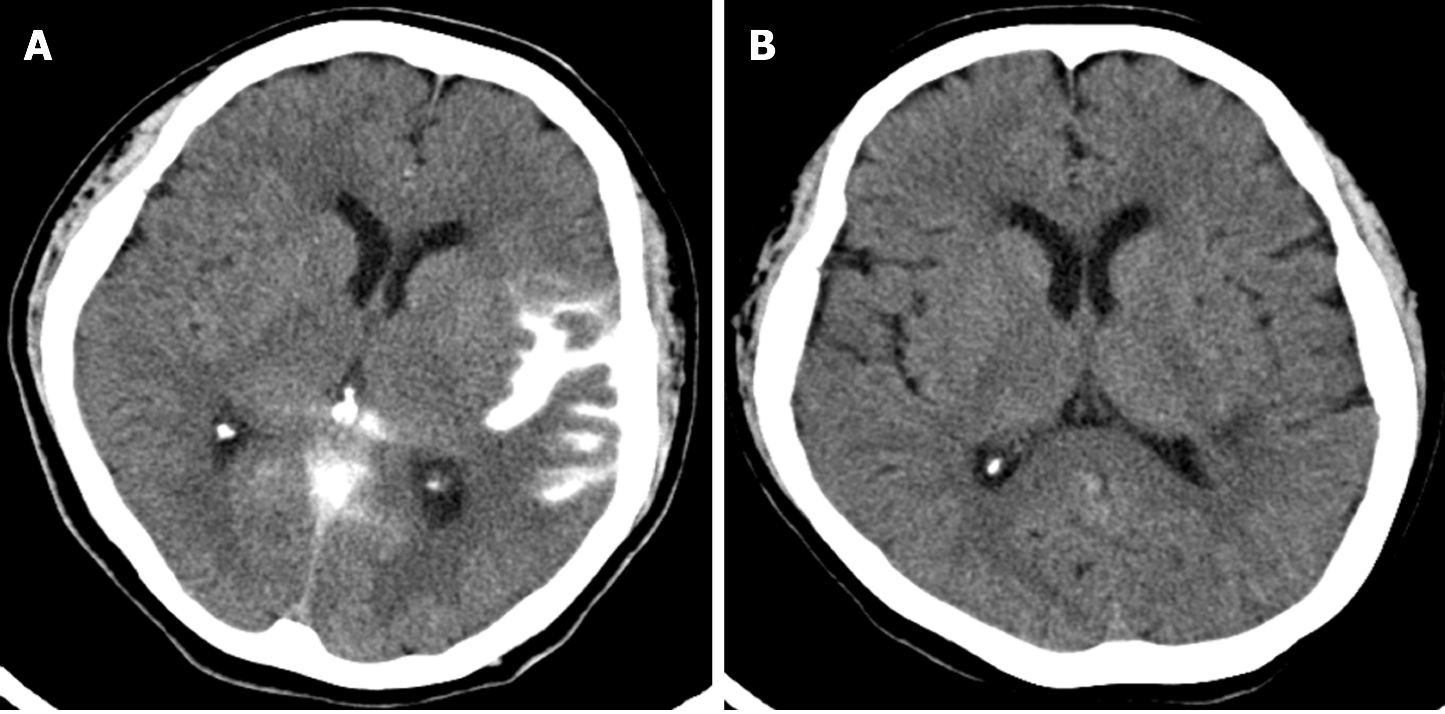
Figure 3 Immediate post-procedure and 12-d follow-up brain computed tomography images.
A: Immediately after the procedure, subarachnoid hemorrhage was identified in the left sylvian cistern on brain computed tomography (CT); B: On the follow-up CT 12 d after the procedure, the subarachnoid hemorrhage was resolved, and a focal low density of infarction was seen in the temporal lobe.
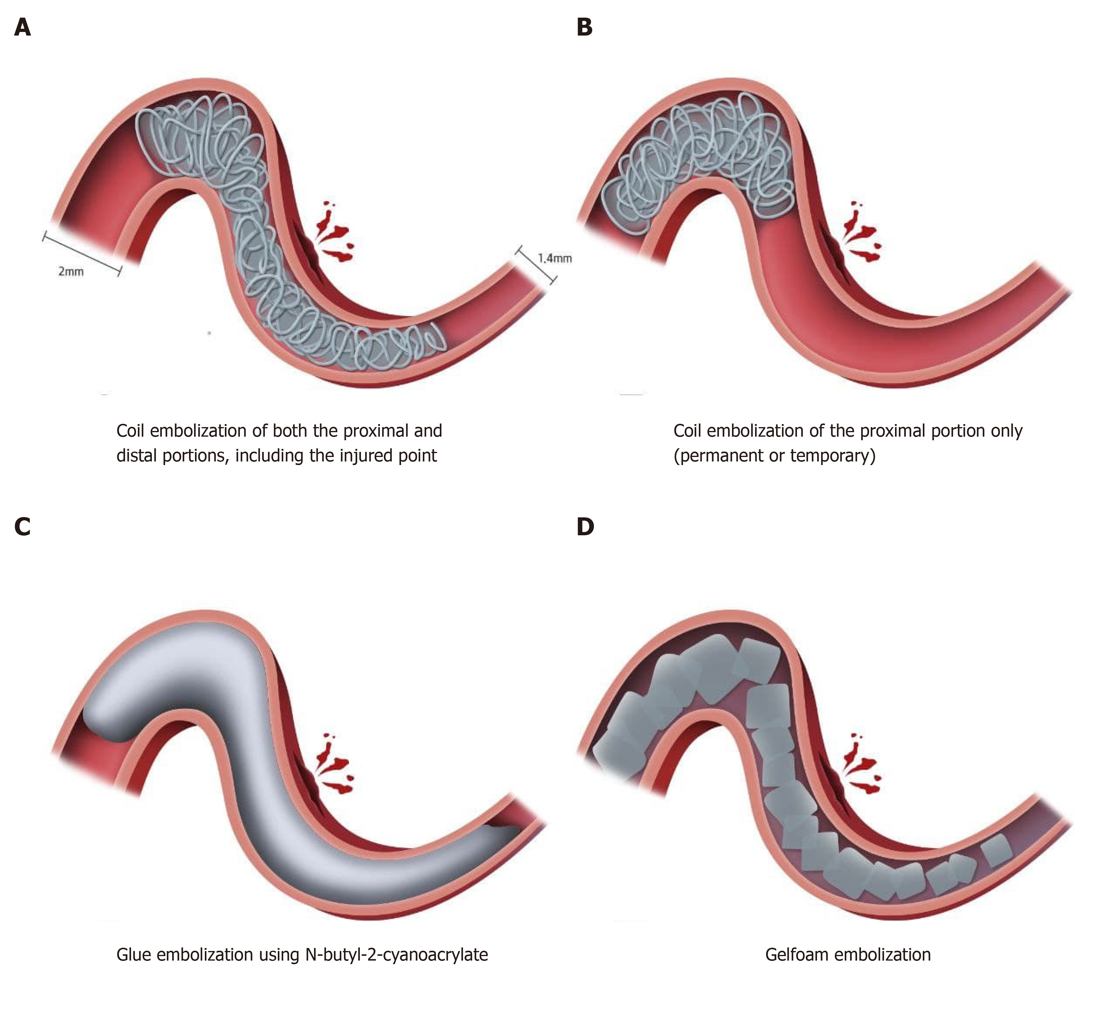
Figure 4 Treatment strategies for rescue embolization of a distal, medium vessel injury during mechanical thrombectomy.
A: Coil embolization of both the proximal and distal portions, including the injured point; B: Coil embolization of the proximal portion only (permanent or temporary); C: Glue embolization using N-butyl-2-cyanoacrylate; D: Gelfoam embolization.
- Citation: Kang JY, Yi KS, Cha SH, Choi CH, Kim Y, Lee J, Cho BS. Gelfoam embolization for distal, medium vessel injury during mechanical thrombectomy in acute stroke: A case report. World J Clin Cases 2021; 9(20): 5668-5674
- URL: https://www.wjgnet.com/2307-8960/full/v9/i20/5668.htm
- DOI: https://dx.doi.org/10.12998/wjcc.v9.i20.5668


