Copyright
©The Author(s) 2021.
World J Clin Cases. Jul 16, 2021; 9(20): 5661-5667
Published online Jul 16, 2021. doi: 10.12998/wjcc.v9.i20.5661
Published online Jul 16, 2021. doi: 10.12998/wjcc.v9.i20.5661
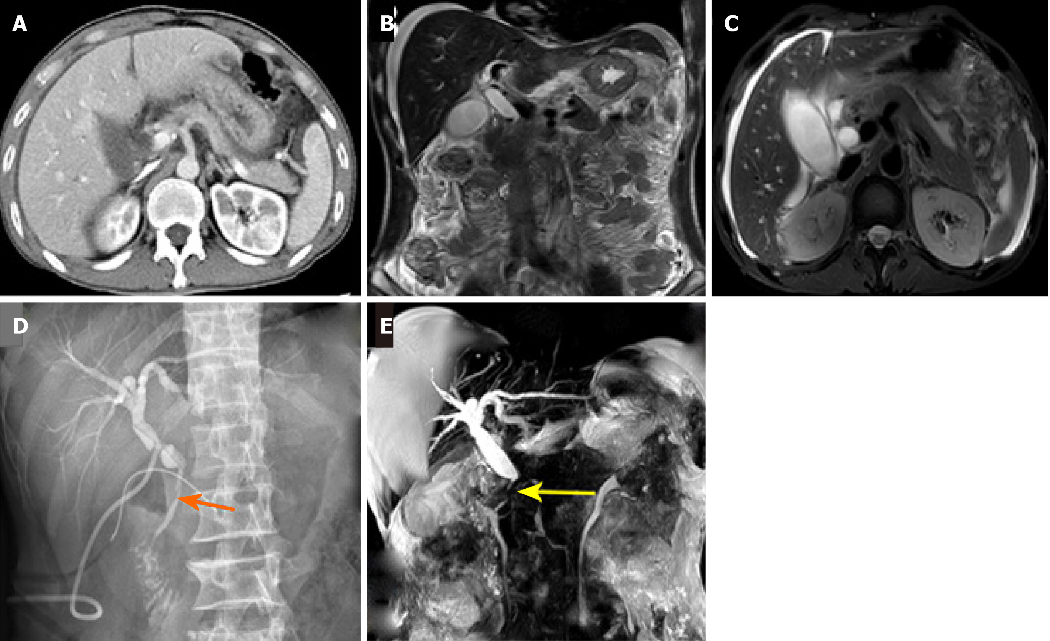
Figure 1 Abdominal computed tomography and magnetic resonance imaging reexamination.
A: Preoperative abdominal computed tomography showed complete intrinsic palpation of the liver, no obvious damage to the abdominal organs and a small amount of perihepatic effusion; B and C: Preoperative magnetic resonance imaging showed good continuity of extrahepatic biliary tract, gallbladder filling, and the causes of perihepatic and pelvic effusion were difficult to determine; D: T-tube angiography at 2 wk postoperatively. The orange arrow indicates the injury site of the lower common bile duct; E: Magnetic resonance cholangiopancreatography (MRCP) was reexamined 2 mo after surgery. The yellow arrow shows the post-traumatic stenosis of the common bile duct. Combined with the results of T-tube angiography at 2 wk postoperatively and MRCP imaging at 2 mo postoperatively, we considered that this was a rare extra-hepatic bile duct injury that had caused mild stenosis of the common bile duct.
- Citation: Zhao J, Dang YL, Lin JM, Hu CH, Yu ZY. Rare isolated extra-hepatic bile duct injury: A case report. World J Clin Cases 2021; 9(20): 5661-5667
- URL: https://www.wjgnet.com/2307-8960/full/v9/i20/5661.htm
- DOI: https://dx.doi.org/10.12998/wjcc.v9.i20.5661


