Copyright
©The Author(s) 2021.
World J Clin Cases. Jul 16, 2021; 9(20): 5647-5654
Published online Jul 16, 2021. doi: 10.12998/wjcc.v9.i20.5647
Published online Jul 16, 2021. doi: 10.12998/wjcc.v9.i20.5647
Figure 1 Enhanced T1-weighted image of both thigh muscles.
A: Coronal section shows enhancement of the right rectus femoris muscle (arrow); B: Transverse section shows heterogeneous enhancement of the vastus lateralis (arrow) and adductor magnus (arrow head) muscles.
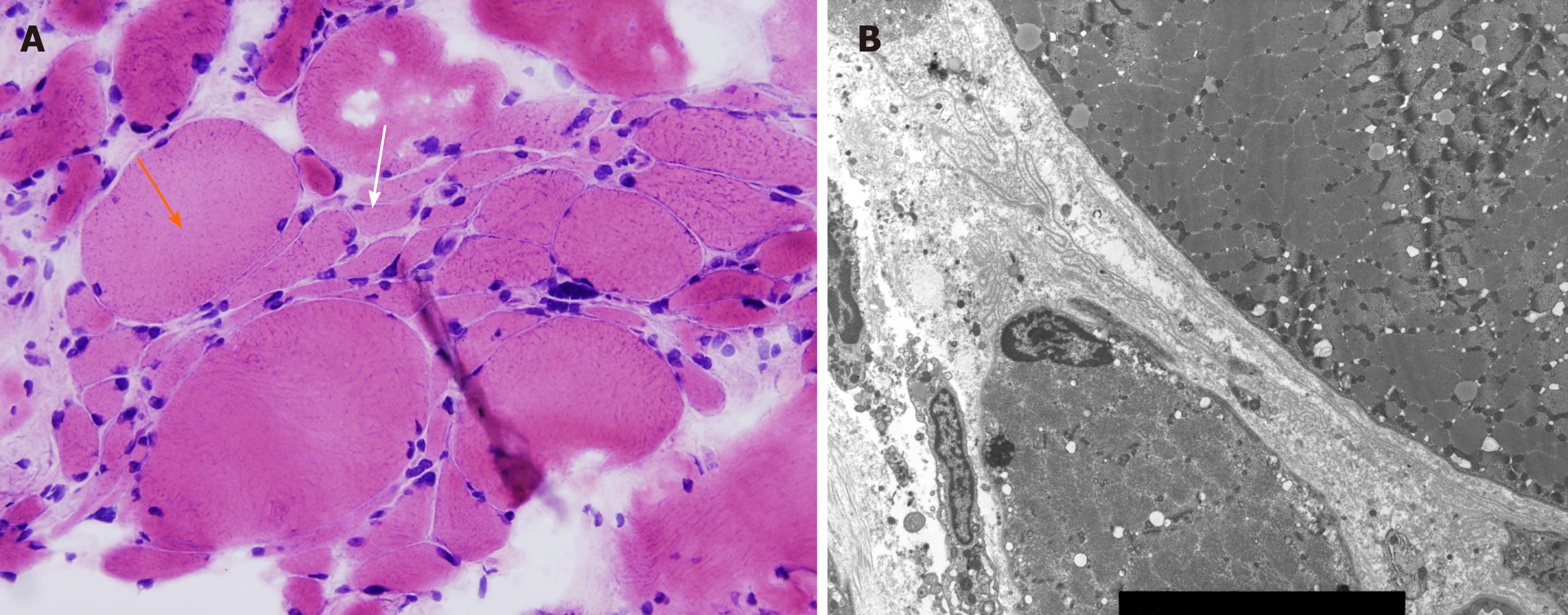
Figure 2 Muscle biopsy of the right vastus lateralis muscle.
A: Light microscopy shows atrophic myofiber (white arrow) and compensatory hypertrophy (orange arrow). Increased endomysial connective tissue is also seen (hematoxylin and eosin, 200 ×); B: Electron microscopy shows atrophic small myofiber with redundant basal lamina.
- Citation: Kim KW, Cho JH. Muscular atrophy and weakness in the lower extremities in Behçet’s disease: A case report and review of literature. World J Clin Cases 2021; 9(20): 5647-5654
- URL: https://www.wjgnet.com/2307-8960/full/v9/i20/5647.htm
- DOI: https://dx.doi.org/10.12998/wjcc.v9.i20.5647


