Copyright
©The Author(s) 2021.
World J Clin Cases. Jul 16, 2021; 9(20): 5594-5604
Published online Jul 16, 2021. doi: 10.12998/wjcc.v9.i20.5594
Published online Jul 16, 2021. doi: 10.12998/wjcc.v9.i20.5594
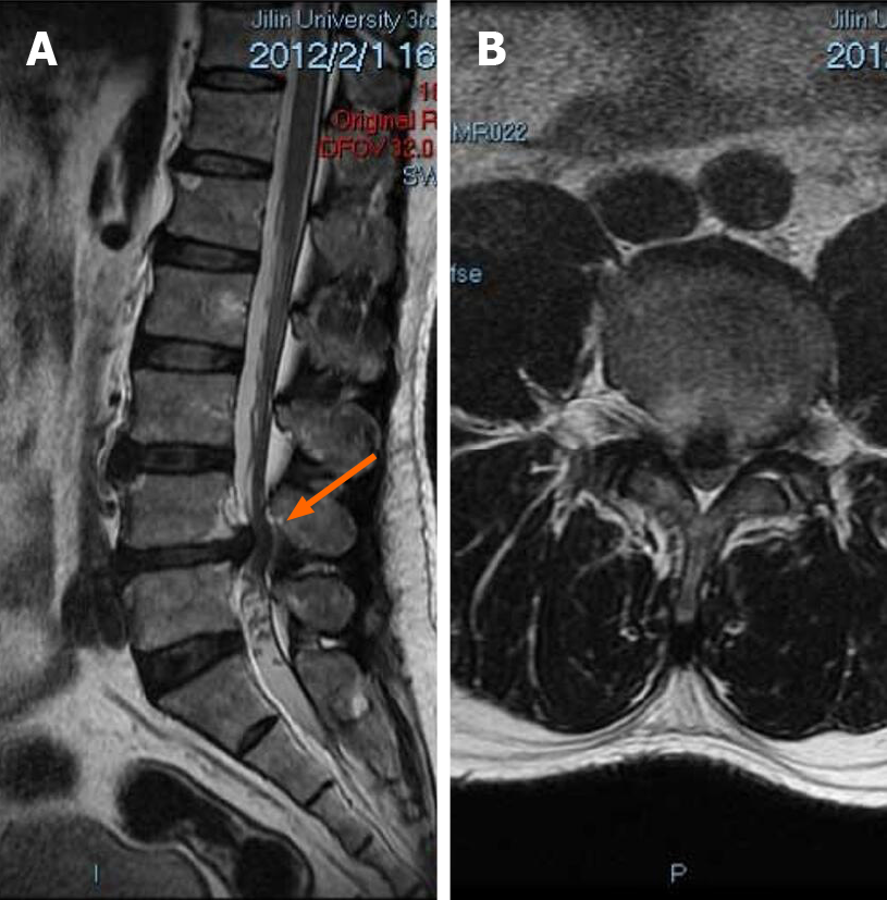
Figure 1 Patient’s first admission preoperative magnetic resonance imaging.
A: Sagittal view of the patient showed a straightening of the physiological curvature of the lumbar spine and a herniated disc at L4-L5 segments (orange arrow); B: Axial scan showed a herniated disc at segments L4-L5, compression of the dura, marked compression of the foramina on both sides and spinal canal stenosis.
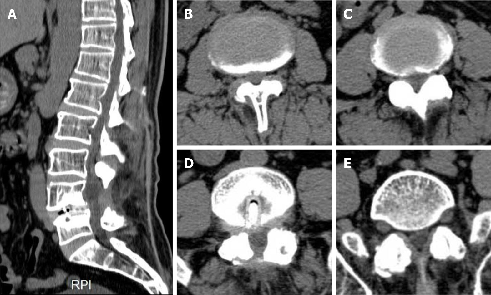
Figure 2 Preoperative computed tomography on the patient’s second admission.
A: Sagittal examination revealed the presence of physiological curvature of the lumbar spine, reduced bone mineral density of all vertebral bodies, irregular margins in some and absence of spinous processes in L4; B-E: L2-L3 (B), L3-L4 (C), L4-L5 (D), L5-S1 (E) cross sections, respectively. Soft tissue density shadowing of multisegmented discs projected towards the periphery. Metal-like structures were found in L4-L5. RPI: Right posterior image.
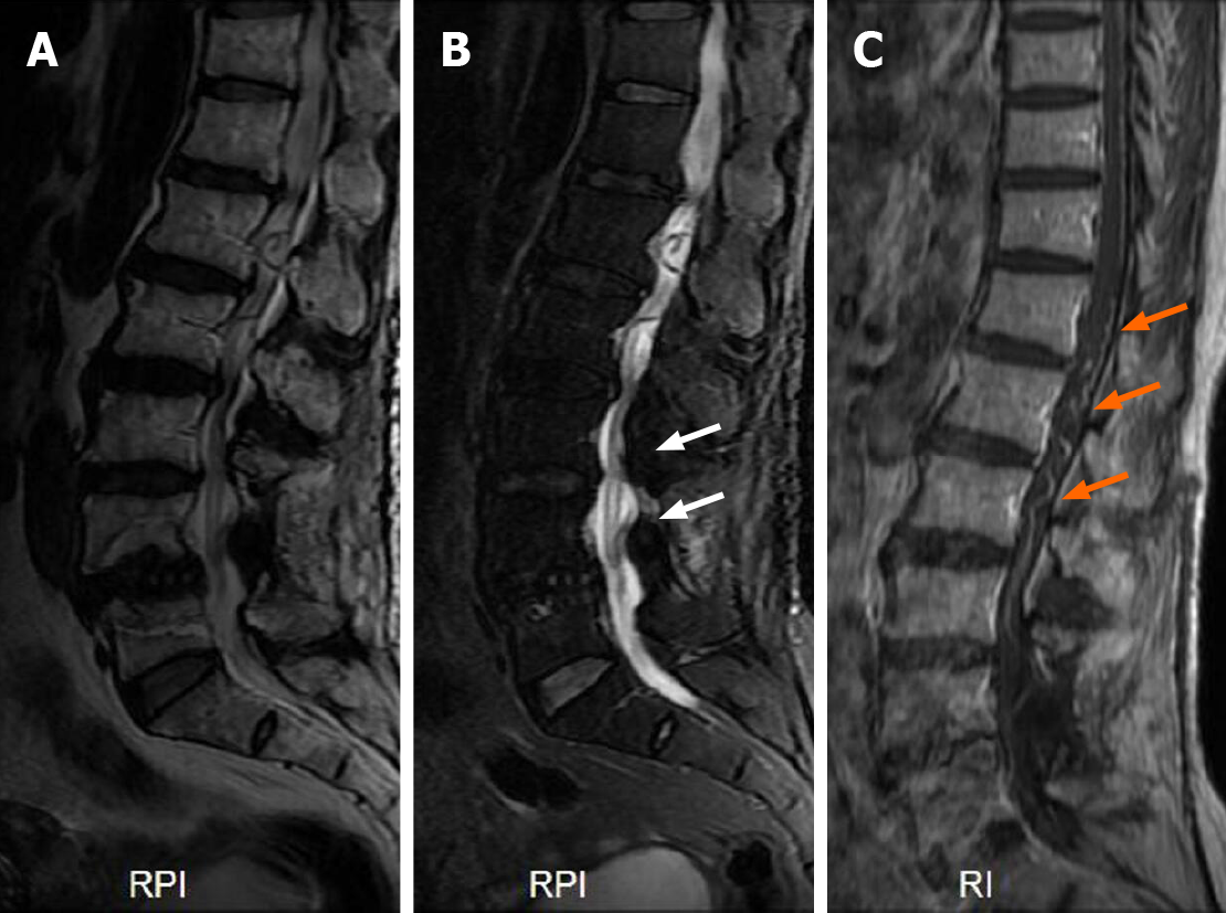
Figure 3 Preoperative magnetic resonance imaging at second admission.
A and B: On T2 image, L4 vertebral body was displaced forward, the edge of vertebral body was irregular, multi segment intervertebral disc protruded backward (A), and high signal shadow was seen in the spinal cord (white arrows) (B); C: Enhanced magnetic resonance imaging showed tortuous dilated vessels in the dorsal side of the spinal cord (orange arrows). RPI: Right posterior image; RI: Right image.
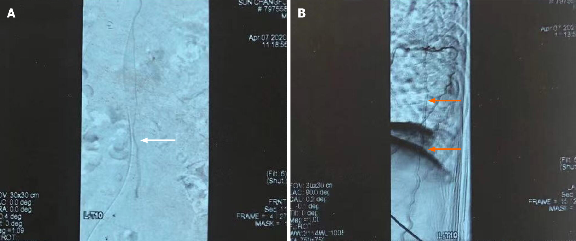
Figure 4 Angiography of spinal cord.
A: Dilated arterialized vessels can be seen in the coronal view (white arrow); B: Sagittal view revealed tortuous dilation and snake-like abnormal veins (orange arrows).
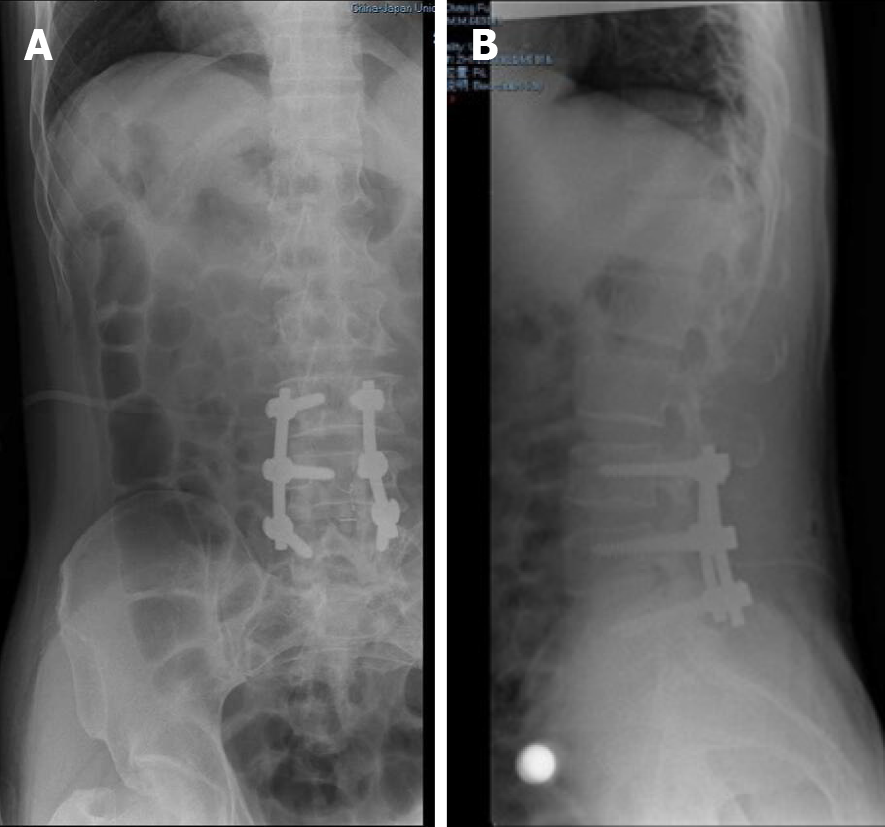
Figure 5 Postoperative X-ray at the patient’s first admission.
A and B: Anteroposterior (A) and lateral (B) radiographs showed that the internal fixative was well positioned in the patient.
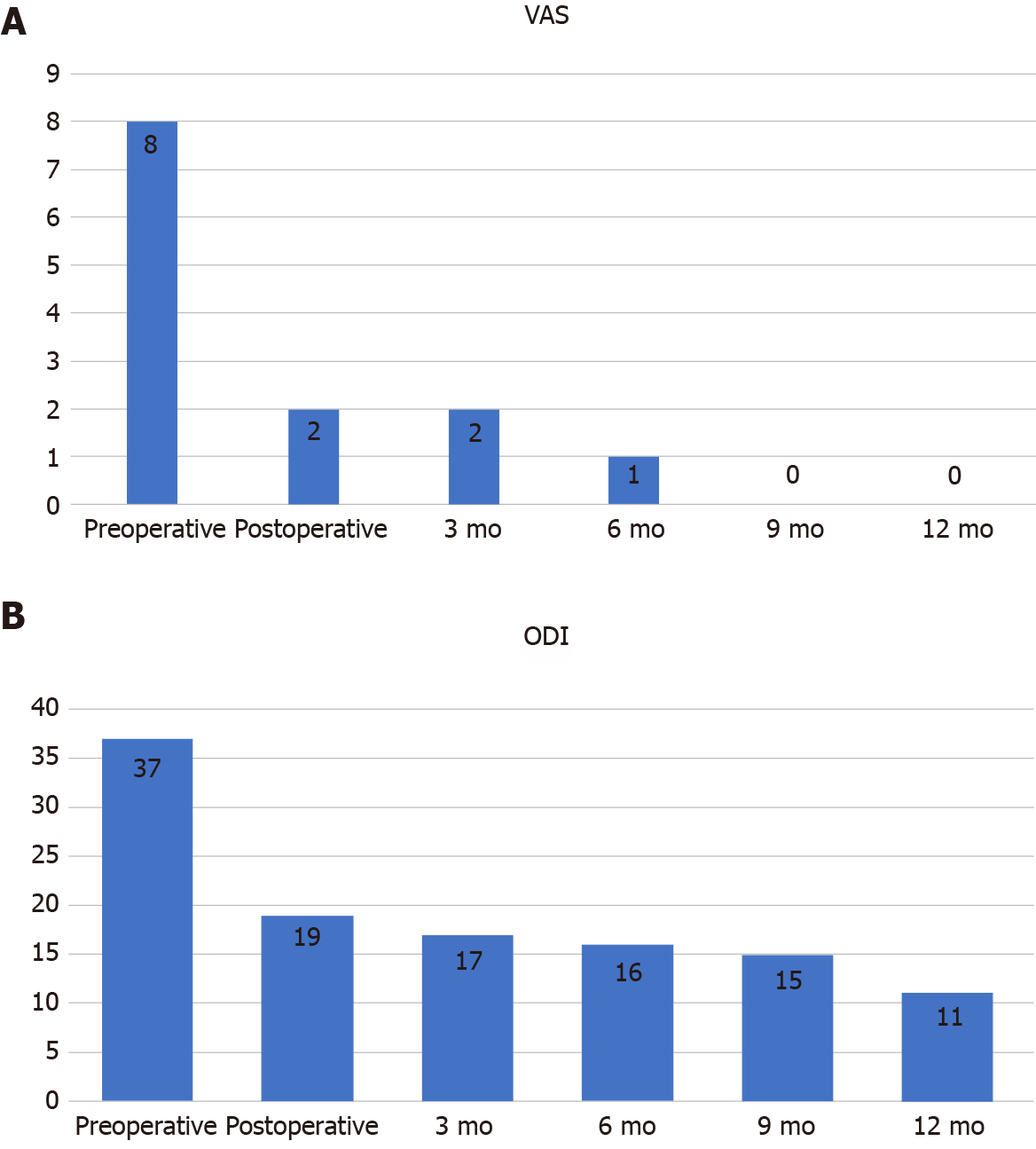
Figure 6 Changes in visual analogue scale scores and Oswestry disability index scores from preoperative to 1 year after surgery.
A: VAS: Visual analogue scale; ODI: Oswestry disability index.
- Citation: Ouyang Y, Qu Y, Dong RP, Kang MY, Yu T, Cheng XL, Zhao JW. Spinal dural arteriovenous fistula 8 years after lumbar discectomy surgery: A case report and review of literature. World J Clin Cases 2021; 9(20): 5594-5604
- URL: https://www.wjgnet.com/2307-8960/full/v9/i20/5594.htm
- DOI: https://dx.doi.org/10.12998/wjcc.v9.i20.5594


