Copyright
©The Author(s) 2021.
World J Clin Cases. Jul 16, 2021; 9(20): 5547-5555
Published online Jul 16, 2021. doi: 10.12998/wjcc.v9.i20.5547
Published online Jul 16, 2021. doi: 10.12998/wjcc.v9.i20.5547
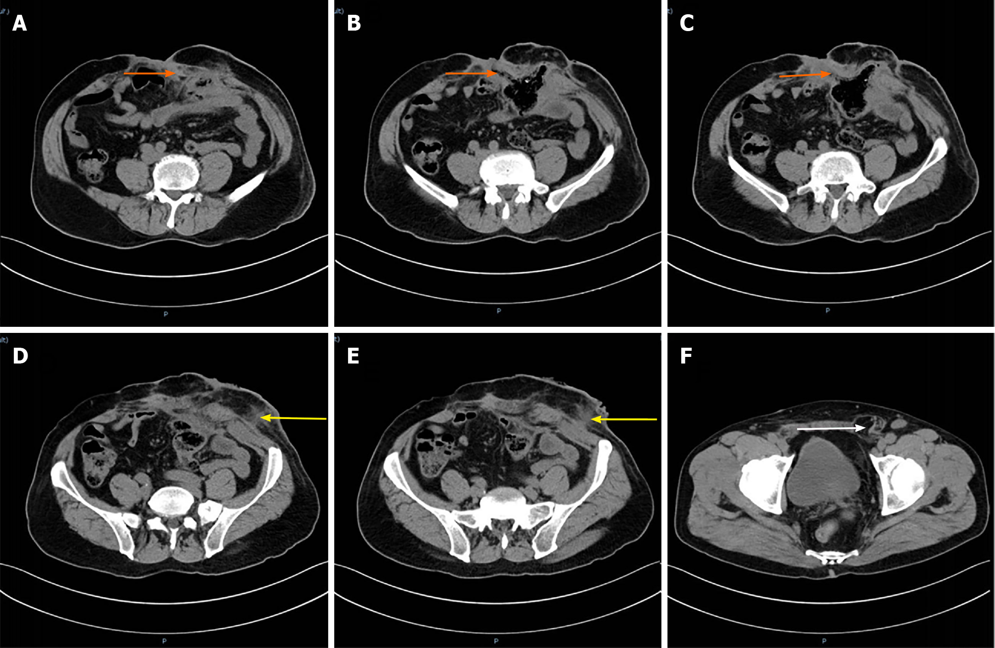
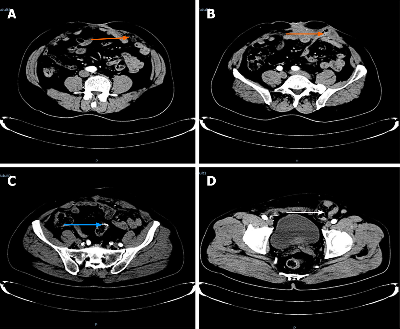
Figure 1 Enhanced computed tomography.
A-C: The orange arrow refers to the sinus formed by the intestinal fistula on the right side of the stoma; D and E: The yellow arrow is the suspicious sinus on the left side of the stoma; F: The white arrow is the left inguinal hernia.
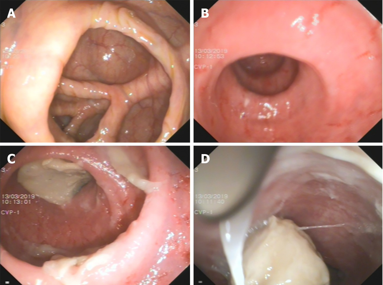
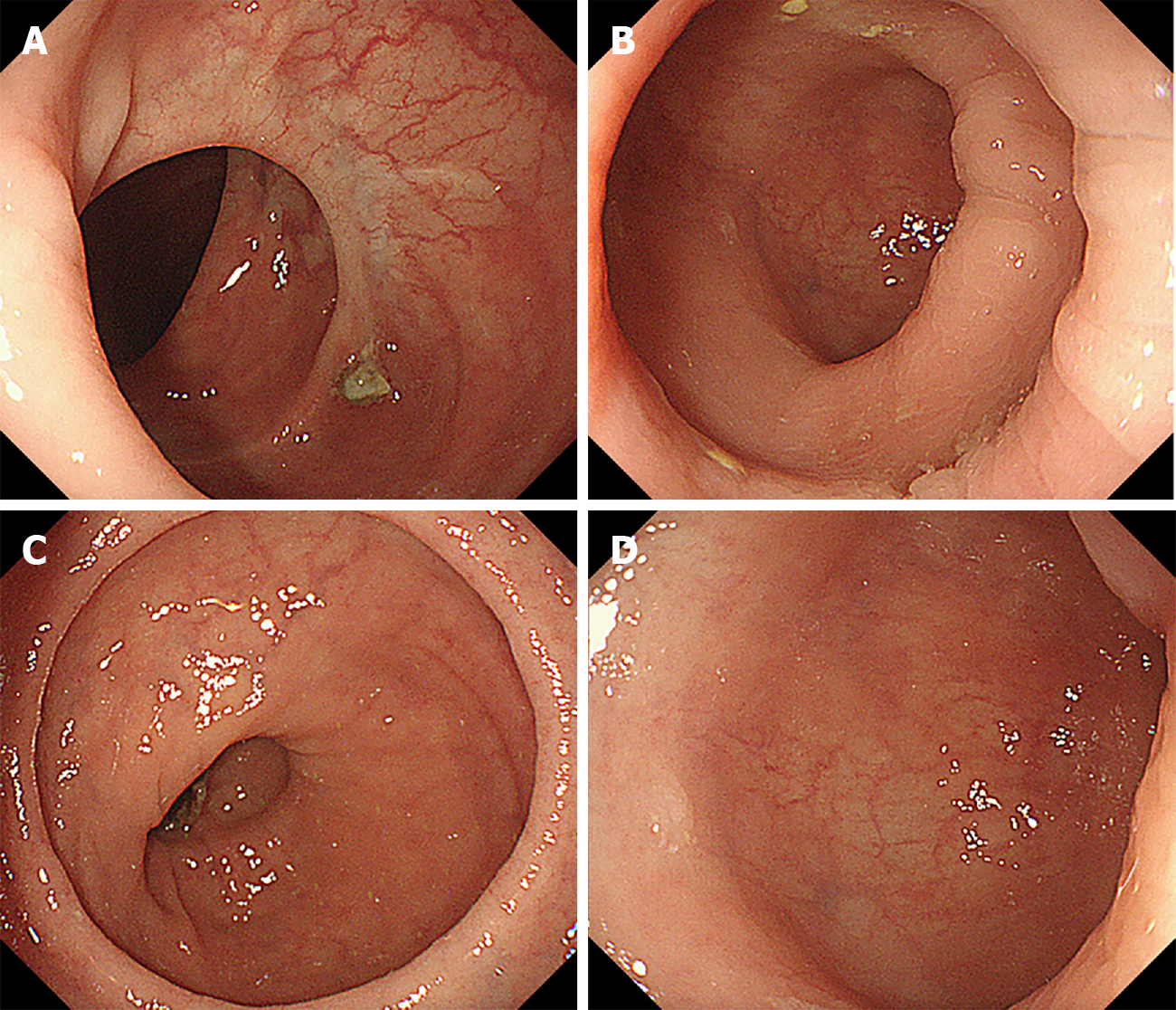
Figure 2 Colonoscopy.
A: The ileocecal region; B: The proximal intestine; C and D: The distal intestine.
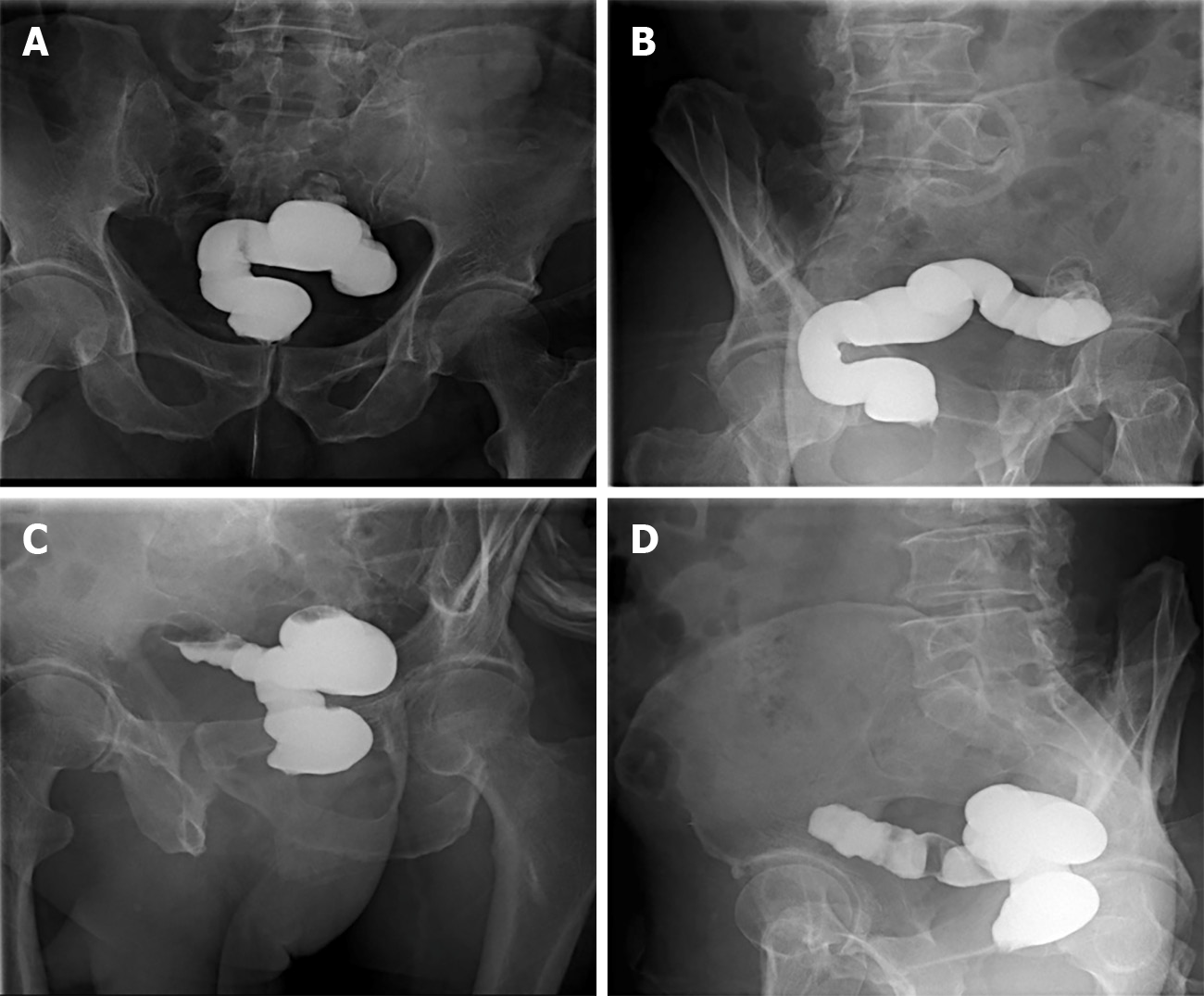
Figure 3 Pelvis films.
A and B: Pelvic anterior films; C and D: Pelvic lateral films.
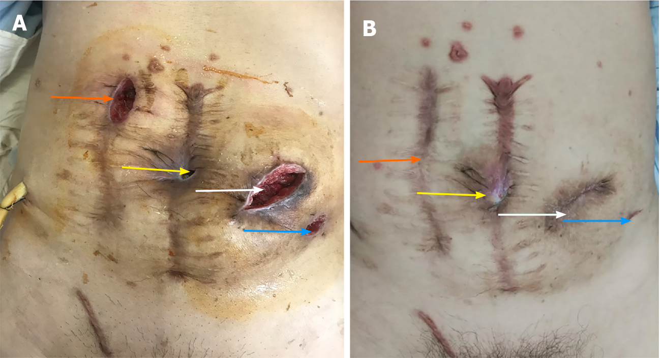
Figure 4 Postoperative wound.
A: The wound on the 6th day after surgery. The orange arrow is the small incision, the yellow arrow is the sinus on the right side of the stoma, the white arrow is the original fistula position, and the blue arrow is the left fistula of the stoma; B: The wound 6 mo after the operation. The yellow arrow is the sinus tract on the right side of the stoma, which has not healed, the white arrow is the location of the original stoma, which is an incisional hernia, and the blue arrow is the left fistula of the original stoma.
Figure 5 Re-examination at 6 mo showed no tumor recurrence.
A and B: The orange arrow is the incisional hernia; C: The blue arrow is the anastomosis; D: The white arrow is the left inguinal hernia, which is larger than before.
Figure 6 A colonoscopy was performed one year after surgery.
A: The proximal end; B and C: The anastomoses; D: The distal end of the anastomosis.
- Citation: Huang W, Chen ZZ, Wei ZQ. Successful reversal of ostomy 13 years after Hartmann procedure in a patient with colon cancer: A case report. World J Clin Cases 2021; 9(20): 5547-5555
- URL: https://www.wjgnet.com/2307-8960/full/v9/i20/5547.htm
- DOI: https://dx.doi.org/10.12998/wjcc.v9.i20.5547


