Copyright
©The Author(s) 2021.
World J Clin Cases. Jul 16, 2021; 9(20): 5540-5546
Published online Jul 16, 2021. doi: 10.12998/wjcc.v9.i20.5540
Published online Jul 16, 2021. doi: 10.12998/wjcc.v9.i20.5540
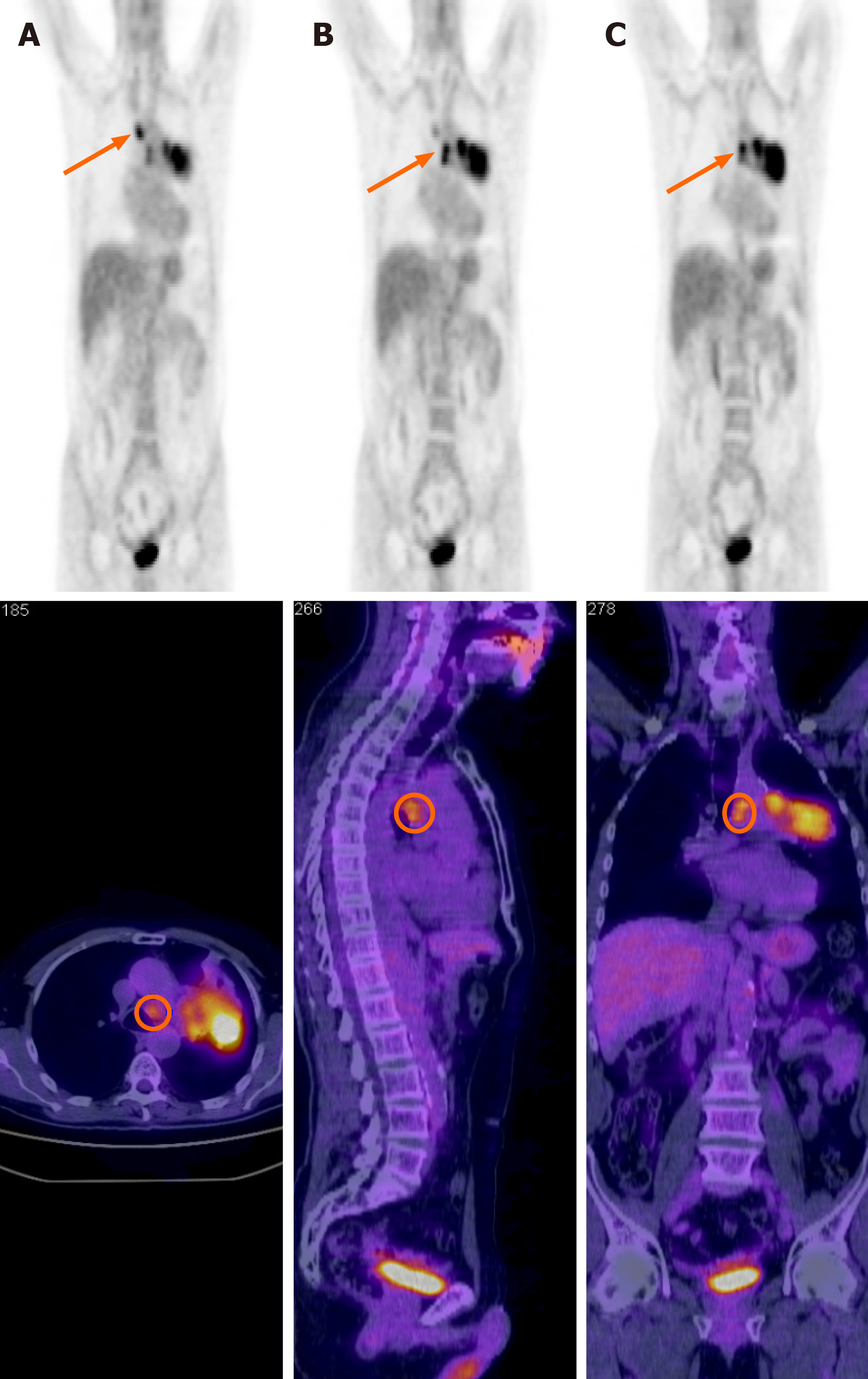
Figure 1 Positron emission tomography/computed tomography staging before the start of chemoradiotherapy treatment.
A: The orange arrow indicates the localization of the involved mediastinal lymph node in station 2R; B: The orange arrow indicates the localization of the involved mediastinal lymph nodes in stations 7L and 4L; C: The orange arrow indicates the localization of the primary tumor with mediastinal infiltration. Radiological staging was cT4 cN3 cM0.
Figure 2 Dose distribution of the patient radiotherapy plan on computed tomography.
The color legend dose-volume histograms in the column on the right illustrates the dose distribution. Prescription dose was 30 Gy/5 daily fractions with a heterogeneous dose escalation of up to 40 Gy inside the primary tumor to simulate brachytherapy dose distribution. Prescription dose was 25 Gy/5 daily fractions with a heterogeneous dose escalation of up to 37.5 Gy inside the nodal tumor. The different colors show the following isodoses: red, 30 Gy; deep blue, 40 Gy; aqua, 37.5 Gy; light blue, 12.5 Gy; green, 25 Gy; pink, 20 Gy; light blue, 12.5 Gy; yellow, 5 Gy.
- Citation: Parisi E, Arpa D, Ghigi G, Micheletti S, Neri E, Tontini L, Pieri M, Romeo A. Complete pathological response in locally advanced non-small-cell lung cancer patient: A case report. World J Clin Cases 2021; 9(20): 5540-5546
- URL: https://www.wjgnet.com/2307-8960/full/v9/i20/5540.htm
- DOI: https://dx.doi.org/10.12998/wjcc.v9.i20.5540


