Copyright
©The Author(s) 2021.
World J Clin Cases. Jul 16, 2021; 9(20): 5535-5539
Published online Jul 16, 2021. doi: 10.12998/wjcc.v9.i20.5535
Published online Jul 16, 2021. doi: 10.12998/wjcc.v9.i20.5535
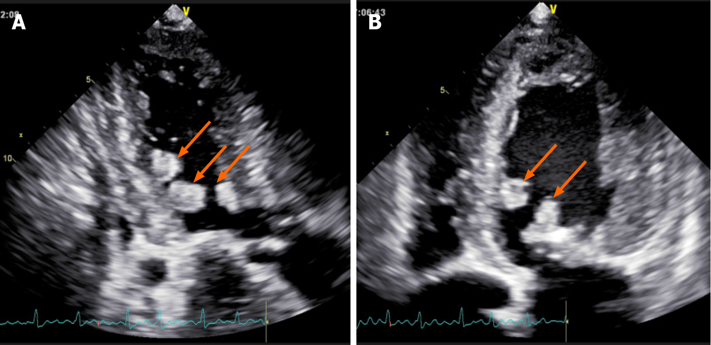
Figure 1 Transesophageal echocardiography demonstrates multiple abnormal masses were in the left ventricle.
A: Apical three chamber view showed abnormal echo masses in the anterior septum and posterior wall of the left ventricle (arrows); B: Apical four chamber echocardiography showed abnormal echo mass in left ventricular posterior septum and mitral leaflet (arrows).
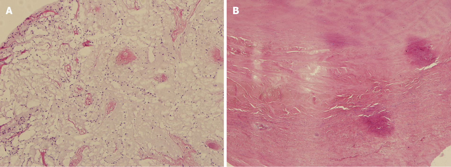
Figure 2 Histopathological findings.
A. The tumor cells were irregular, surrounded by empty halos, and scattered with sparse stroma; B: The valve specimens manifested fibrous tissue hyperplasia, hyaline degeneration, and mucoid degeneration.
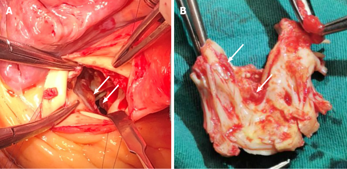
Figure 3 Intra-operative finding.
A: Multiple masses were found in the left ventricular outflow tract (arrows); B: Multiple masses were found in the anterior mitral valve (arrows).
- Citation: Liu SZ, Hong Y, Huang KL, Li XP. Multiple left ventricular myxomas combined with severe rheumatic valvular lesions: A case report. World J Clin Cases 2021; 9(20): 5535-5539
- URL: https://www.wjgnet.com/2307-8960/full/v9/i20/5535.htm
- DOI: https://dx.doi.org/10.12998/wjcc.v9.i20.5535


