Copyright
©The Author(s) 2021.
World J Clin Cases. Jan 16, 2021; 9(2): 445-456
Published online Jan 16, 2021. doi: 10.12998/wjcc.v9.i2.445
Published online Jan 16, 2021. doi: 10.12998/wjcc.v9.i2.445
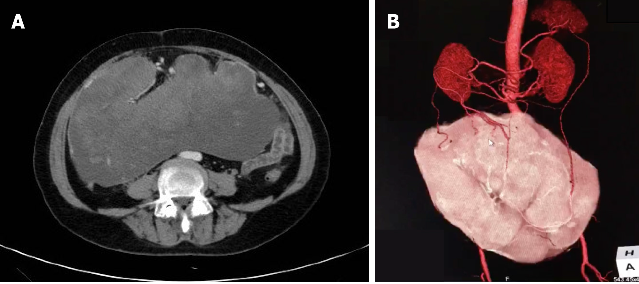
Figure 1 Imaging of the abdominal mass.
A: Contrast-enhanced abdominal computed tomography (CT) scan showing a huge mass measuring 25.4 cm × 23.0 cm with mixed density and heterogeneous enhancement (arrow); B: CT 3D reconstruction showing that the feeding arteries were from the splenic artery, right colic artery, and middle colic artery (arrows).
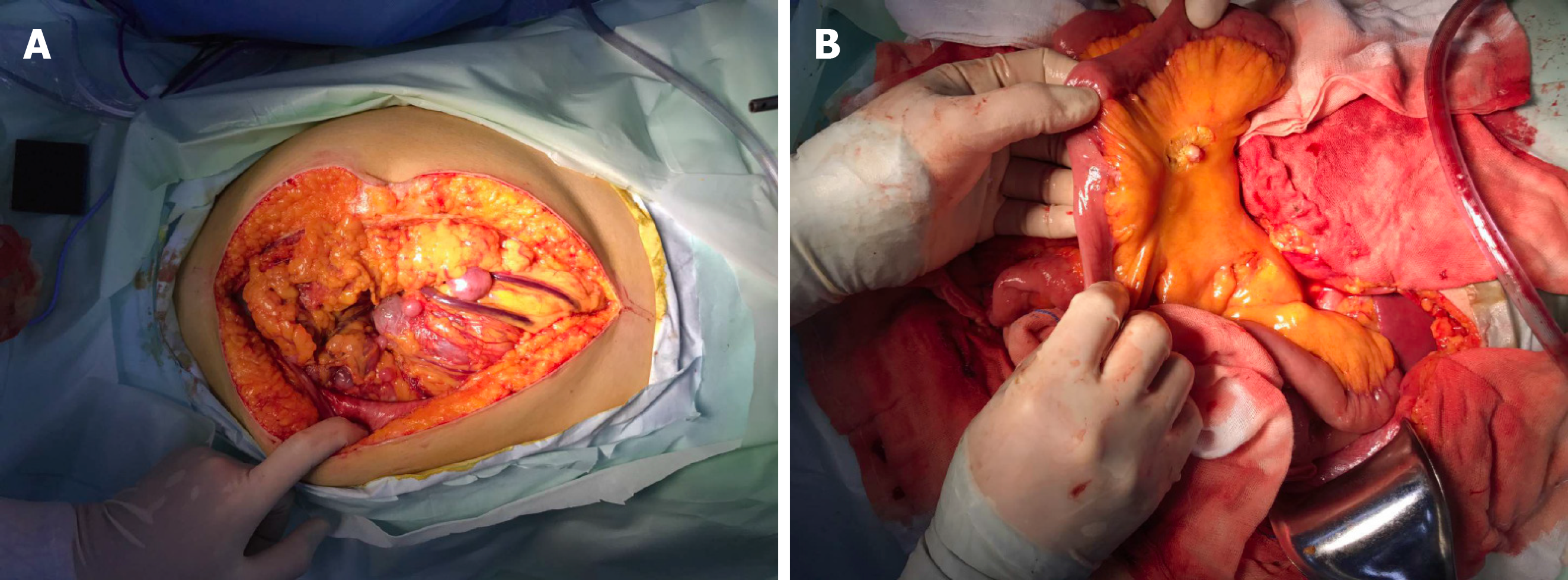
Figure 2 Intraoperative external phase of the abdominal mass.
A: During the operation, a tumor originating from the greater omentum was detected, which occupied most of the space in the abdominal cavity; B: The tumor was highly invasive, and several implant nodules were identified on the intestinal mesentery and greater omentum. A mesenteric implant is shown (arrow).
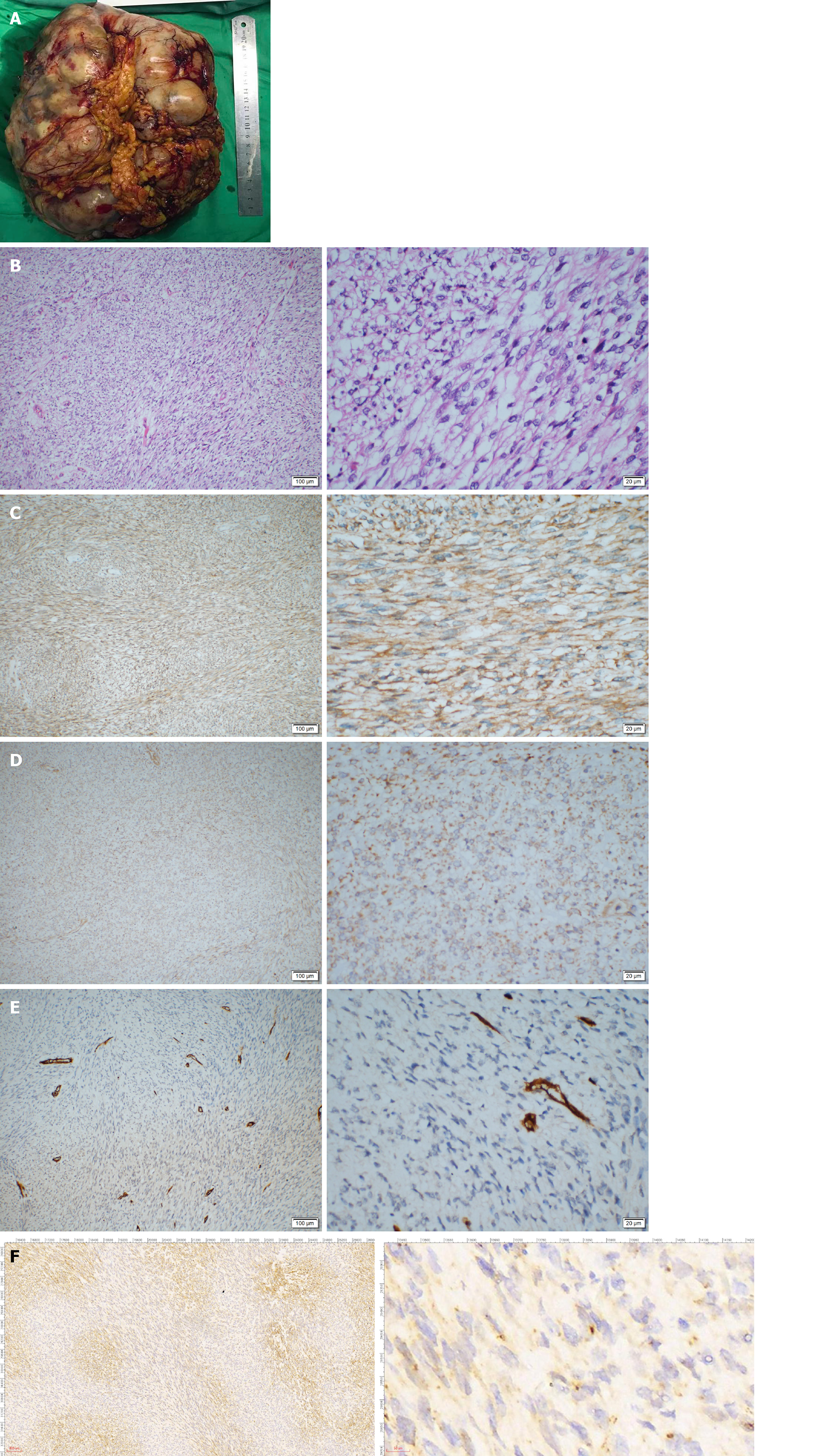
Figure 3 Pathological examination of the tumor.
A: The gross morphology of the resected tumor specimen was huge (27 cm × 21 cm × 9 cm) with necrosis and hemorrhage on the surface; B: Pathological examination showed hypercellularity with spindle cells, and a high mitotic activity with a rate of 30/10 high power field (HPF); C and D: On immunohistochemistry staining, BCL-2 and DOG-1 were weakly positive; E and F: CD34 and STAT6 were negative.
Figure 4 Gene sequencing was performed using the paraffin-embedded tumor section.
A-F: Four exons of c-KIT including 9 (A), 11 (B), 13 (C), and 17 (D); and 2 exons of PDGFRA including 12 (E) and 18 (F) were sequenced using the Sanger method. The results revealed negative gene mutation in all the exons.
- Citation: Guo YC, Yao LY, Tian ZS, Shi B, Liu Y, Wang YY. Malignant solitary fibrous tumor of the greater omentum: A case report and review of literature. World J Clin Cases 2021; 9(2): 445-456
- URL: https://www.wjgnet.com/2307-8960/full/v9/i2/445.htm
- DOI: https://dx.doi.org/10.12998/wjcc.v9.i2.445


