Copyright
©The Author(s) 2021.
World J Clin Cases. Jul 6, 2021; 9(19): 5325-5331
Published online Jul 6, 2021. doi: 10.12998/wjcc.v9.i19.5325
Published online Jul 6, 2021. doi: 10.12998/wjcc.v9.i19.5325
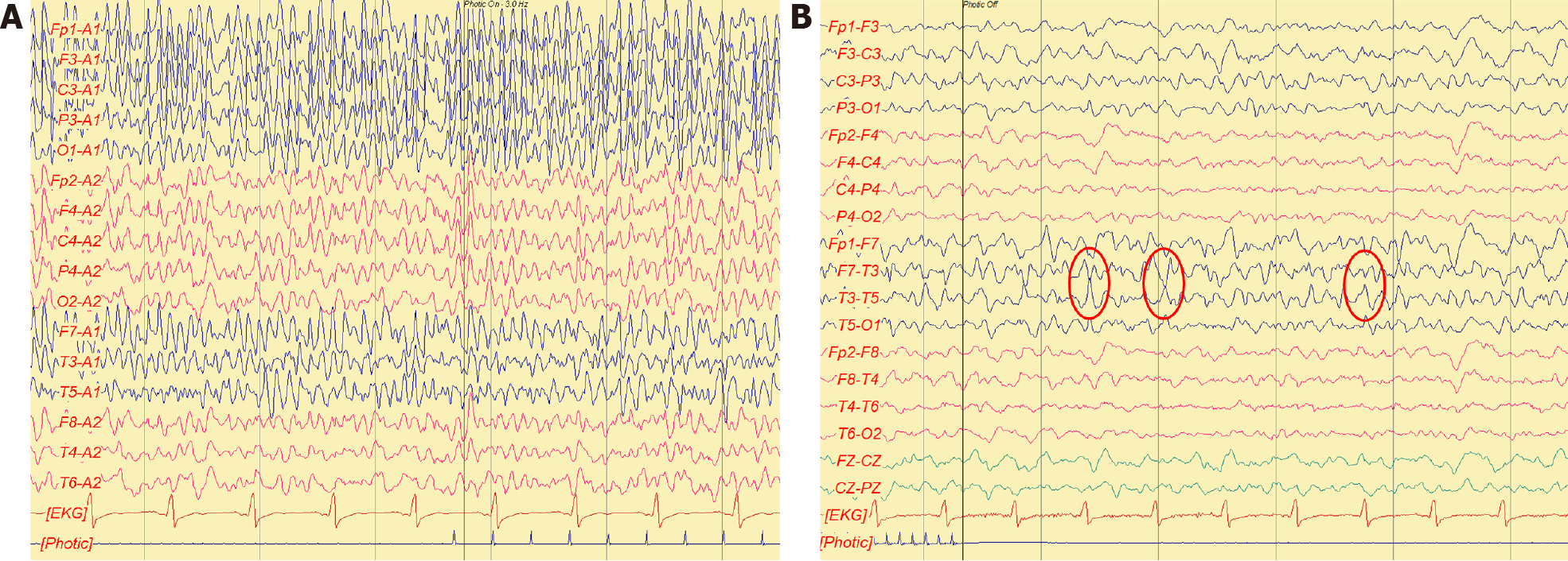
Figure 1 Electroencephalogram.
A: A1–A2 montage of electroencephalogram (EEG), frequent generalized paroxysmal sharp waves with maximal amplitude in the left hemisphere were noticed; B: Double banana montage of EEG, focal paroxysmal sharp waves with phase reverse at T3, which are suggestive of focal epileptogenicity in the left temporal region.
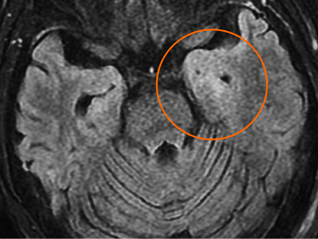
Figure 2 Brain magnetic resonance imaging.
- Citation: Yang CY, Tsai ST. Glutamic acid decarboxylase 65-positive autoimmune encephalitis presenting with gelastic seizure, responsive to steroid: A case report. World J Clin Cases 2021; 9(19): 5325-5331
- URL: https://www.wjgnet.com/2307-8960/full/v9/i19/5325.htm
- DOI: https://dx.doi.org/10.12998/wjcc.v9.i19.5325


