Copyright
©The Author(s) 2021.
World J Clin Cases. Jul 6, 2021; 9(19): 5313-5318
Published online Jul 6, 2021. doi: 10.12998/wjcc.v9.i19.5313
Published online Jul 6, 2021. doi: 10.12998/wjcc.v9.i19.5313
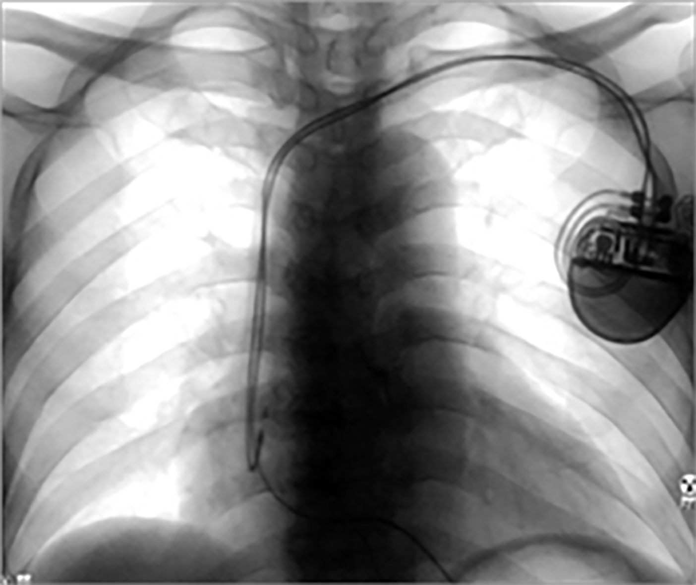
Figure 1 An AP view on first videofluoroscopic swallowing study.
The picture showed implanted cardiac pacemaker and its two leads toward right atrium and right ventricle.
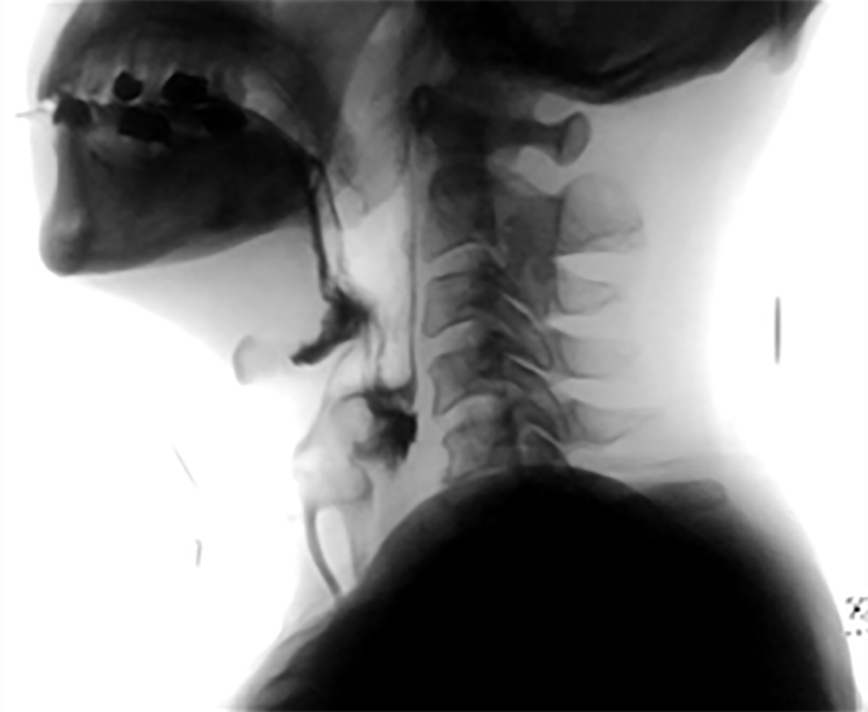
Figure 2 A lateral view on first videofluoroscopic swallowing study.
The picture showed aspirated thin water to trachea.
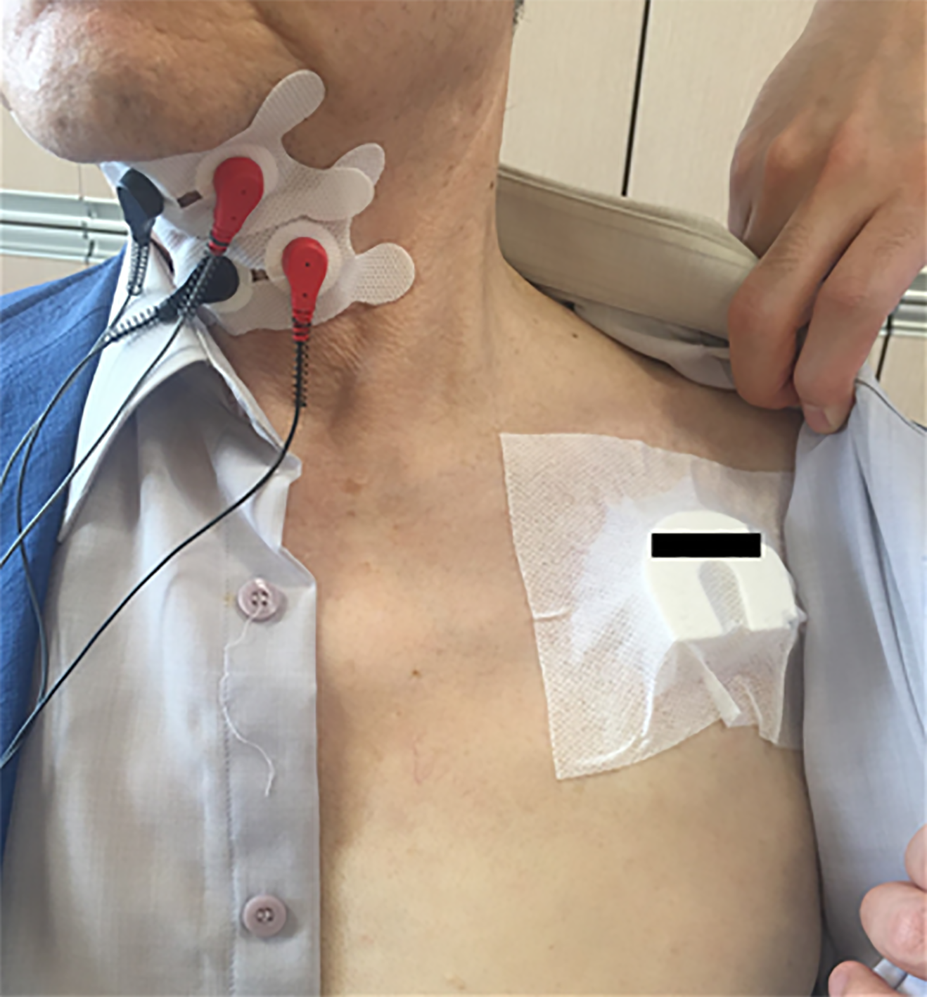
Figure 3 Neuromuscular electrical stimulation treatment with clinical magnet application.
- Citation: Kim M, Park JK, Lee JY, Kim MJ. Neuromuscular electrical stimulation for a dysphagic stroke patient with cardiac pacemaker using magnet mode change: A case report. World J Clin Cases 2021; 9(19): 5313-5318
- URL: https://www.wjgnet.com/2307-8960/full/v9/i19/5313.htm
- DOI: https://dx.doi.org/10.12998/wjcc.v9.i19.5313


