Copyright
©The Author(s) 2021.
World J Clin Cases. Jul 6, 2021; 9(19): 5302-5312
Published online Jul 6, 2021. doi: 10.12998/wjcc.v9.i19.5302
Published online Jul 6, 2021. doi: 10.12998/wjcc.v9.i19.5302
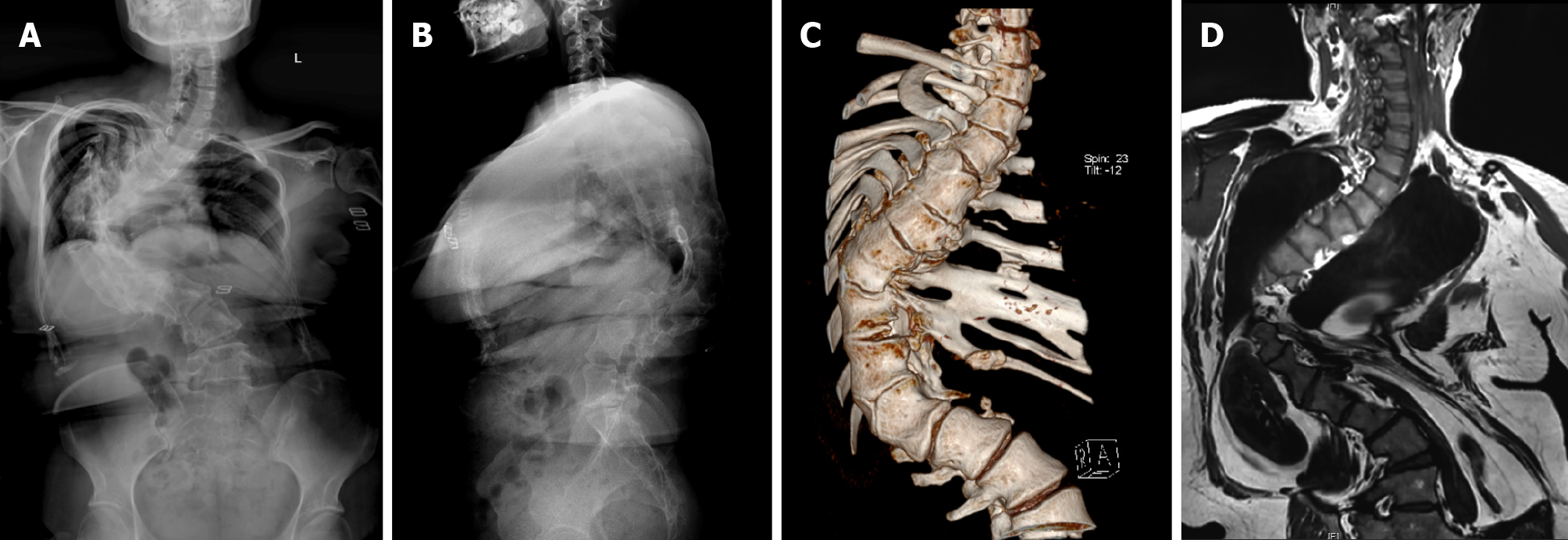
Figure 1 Preoperative imaging examination.
A: X-ray of the scoliosis - AP view; B: X-ray of the scoliosis - Perfil view; C: Computed tomography three-dimensional reconstruction of the patient; D: Magnetic resonance imaging of the patient’s entire spine.
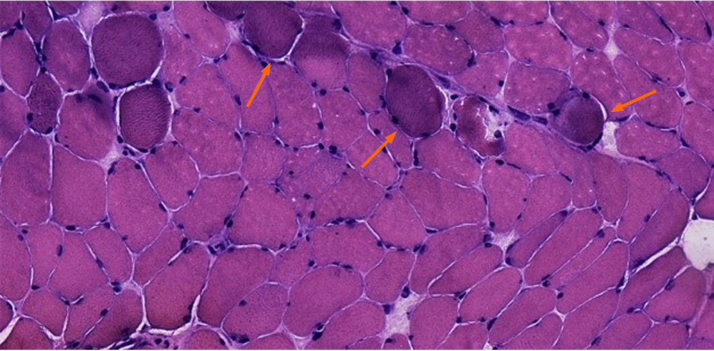
Figure 2 Multifidus biopsy results of hematoxylin and eosin staining.
A few muscles were slightly reduced in size, round in shape, bundles scattered, widened fiber gap and shrink nuclei, with occasional muscle fissure and connective tissue hyperplasia (orange arrows) (hematoxylin & eosin staining, × 40).
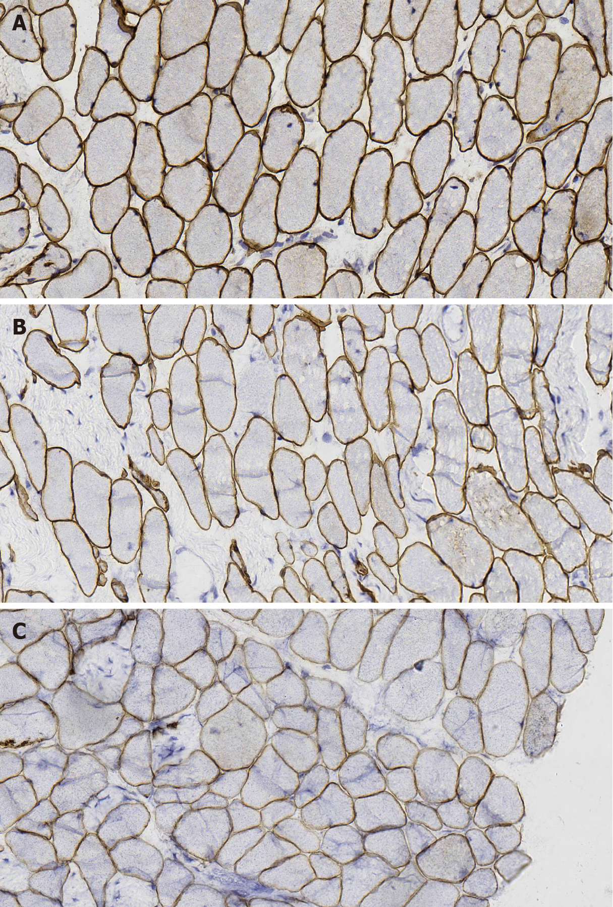
Figure 3 Multifidus biopsy results of dystrophin.
A: Positive dystrophin 1 staining of sarcolemma (immunohistochemistry [IHC]: dystrophin-1 staining, × 40); B: Partial sarcolemma dystrophin 2 staining was uneven (IHC: dystrophin-2 staining, × 40); C: Partial muscle sarcolemma dystrophin 3 stained unevenly (IHC: dystrophin-3, × 40).
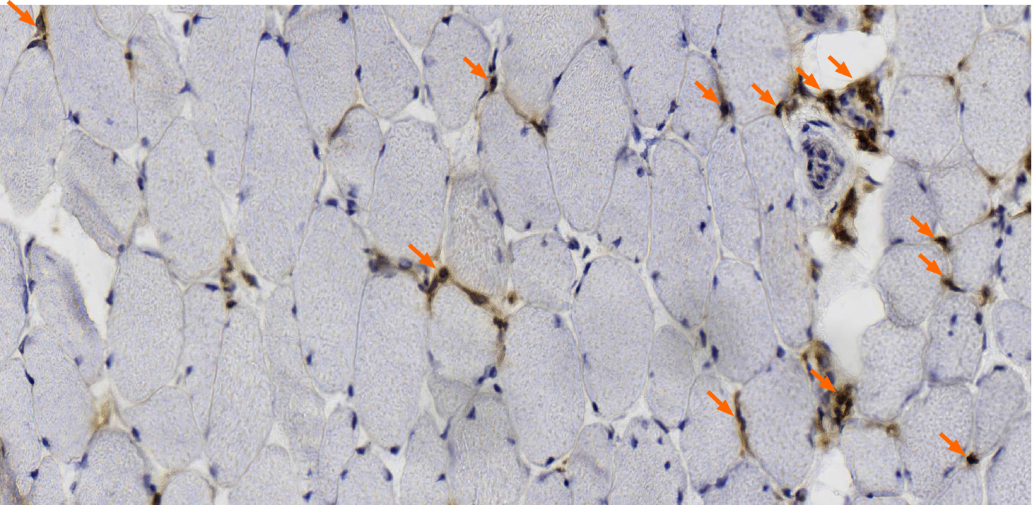
Figure 4 Multifidus biopsy results of cluster of differentiation 4.
A few cluster of differentiation 4 (CD4)-positive staining were seen in the wall and stroma of focal small blood vessels (orange arrows) (CD4 staining, × 40).
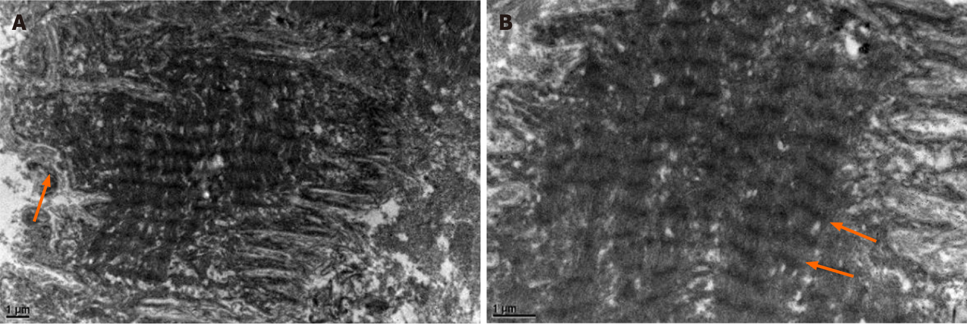
Figure 5 Electron microscopy results.
A: Some sarcolemma is corrugated, and extracellular matrix arrangement is slightly disordered (orange arrows) (× 6000); B: The inner myofibril arrangement is orderly, but band A is unclear (orange arrows) (× 15000).
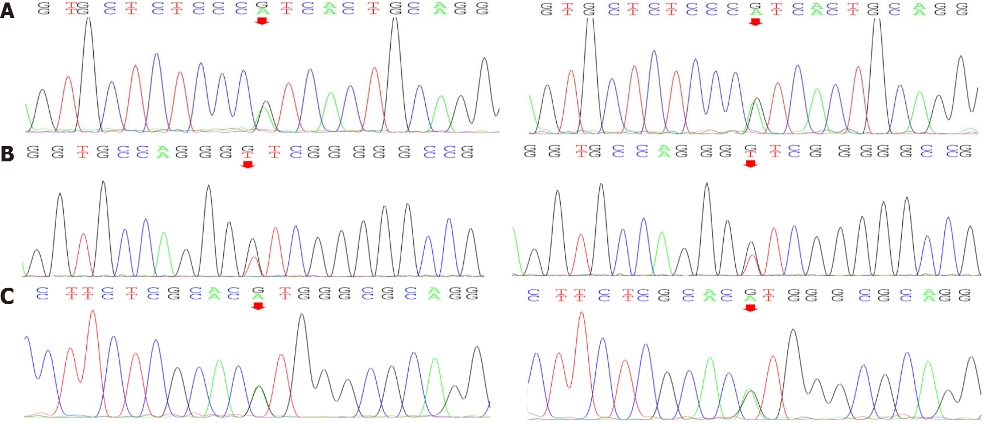
Figure 6 Gene mutations in COL6A1, COL6A2.
A: Sequencing of collagen type VI alpha 1 chain (COL6A1) gene revealed splicing mutations; B: Sequencing of COL6A2 gene revealed missense mutations; C: Sequencing of COL6A2 gene revealed splicing mutations.
Figure 7 Collagen VI immunohistochemistry.
A: This patient muscle membrane stained shallow, broaden and discontinuous (immunohistochemistry [IHC]: collagen type VI alpha (COL6A staining, × 40); B: Another myopathy patient (non-scoliosis) matched by age and sex, staining continuous and dense (IHC: COL6A staining, × 40).
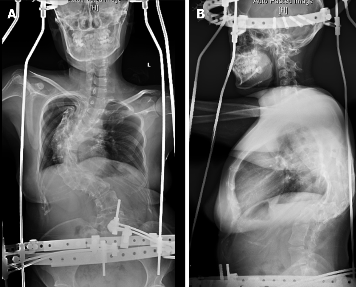
Figure 8 Imaging of halo-pelvic distraction.
A: Postoperative X-ray of the spine (AP view); B: Postoperative X-ray of the spine (Perfil view).
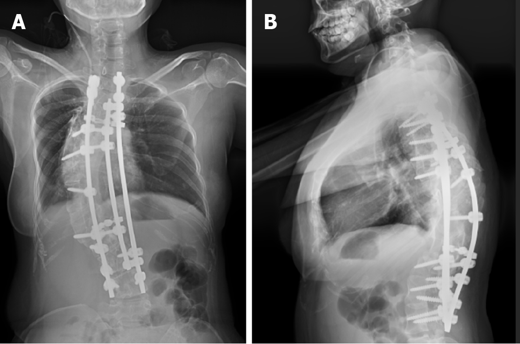
Figure 9 Postoperative imaging examination.
A: Postoperative X-ray of the spine (AP view); B: Postoperative X-ray of the spine (Perfil view).
- Citation: Li JY, Liu SZ, Zheng DF, Zhang YS, Yu M. Collagen VI-related myopathy with scoliosis alone: A case report and literature review . World J Clin Cases 2021; 9(19): 5302-5312
- URL: https://www.wjgnet.com/2307-8960/full/v9/i19/5302.htm
- DOI: https://dx.doi.org/10.12998/wjcc.v9.i19.5302


