Copyright
©The Author(s) 2021.
World J Clin Cases. Jul 6, 2021; 9(19): 5294-5301
Published online Jul 6, 2021. doi: 10.12998/wjcc.v9.i19.5294
Published online Jul 6, 2021. doi: 10.12998/wjcc.v9.i19.5294
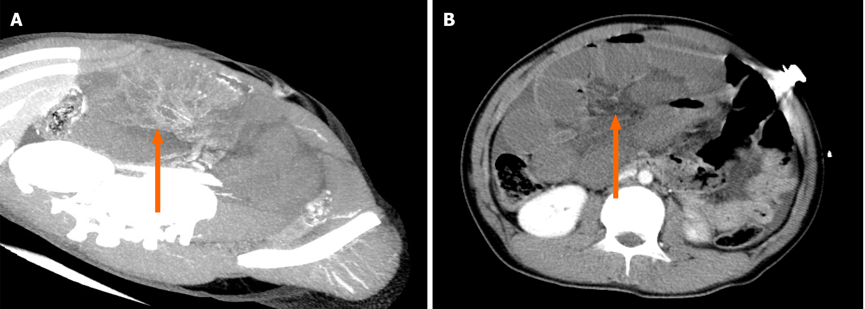
Figure 1 Computed tomography revealed a suspected internal hernia, extensive small intestinal obstruction, and massive effusion in the abdominal and pelvic cavity.
A: Abdominal cavity; B: Pelvic cavity.
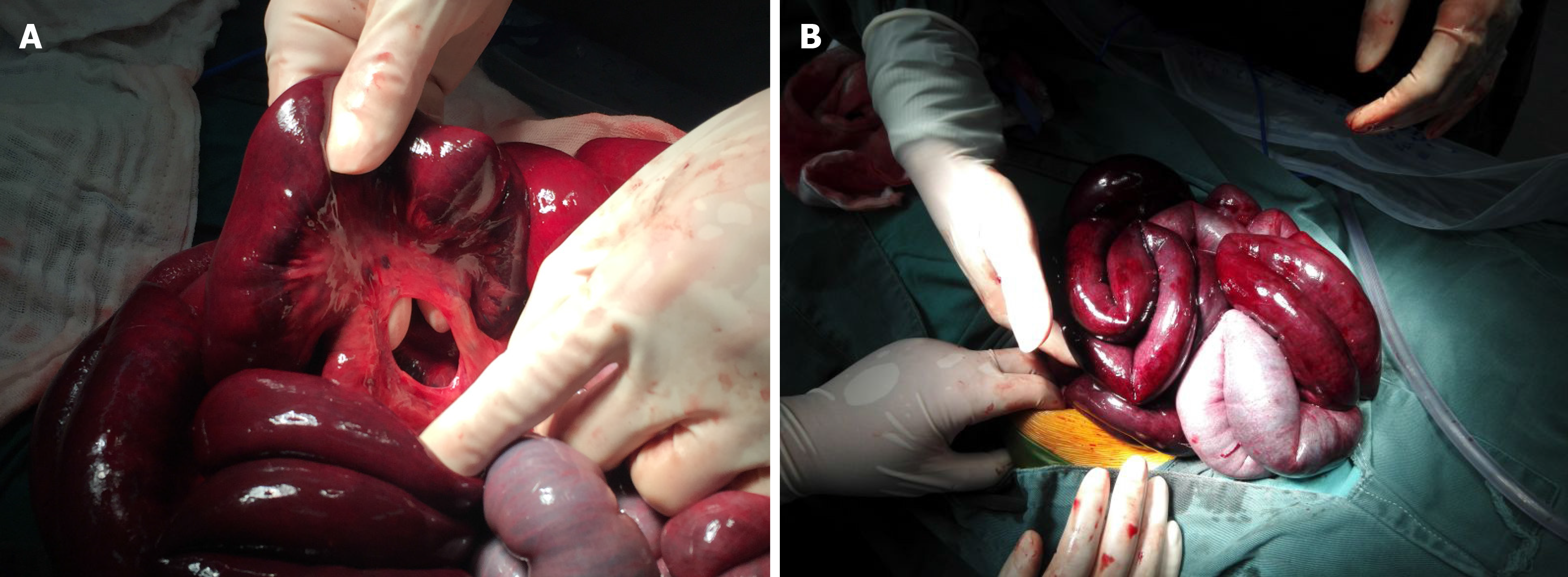
Figure 2 Small intestine images.
A: Small mesenteric defects approximately 3.5 cm in diameter near the ileocecal valve; B: The length of the herniated small intestine was approximately 1.8 m.
- Citation: Zheng XX, Wang KP, Xiang CM, Jin C, Zhu PF, Jiang T, Li SH, Lin YZ. Intestinal gangrene secondary to congenital transmesenteric hernia in a child misdiagnosed with gastrointestinal bleeding: A case report. World J Clin Cases 2021; 9(19): 5294-5301
- URL: https://www.wjgnet.com/2307-8960/full/v9/i19/5294.htm
- DOI: https://dx.doi.org/10.12998/wjcc.v9.i19.5294


