Copyright
©The Author(s) 2021.
World J Clin Cases. Jul 6, 2021; 9(19): 5280-5286
Published online Jul 6, 2021. doi: 10.12998/wjcc.v9.i19.5280
Published online Jul 6, 2021. doi: 10.12998/wjcc.v9.i19.5280
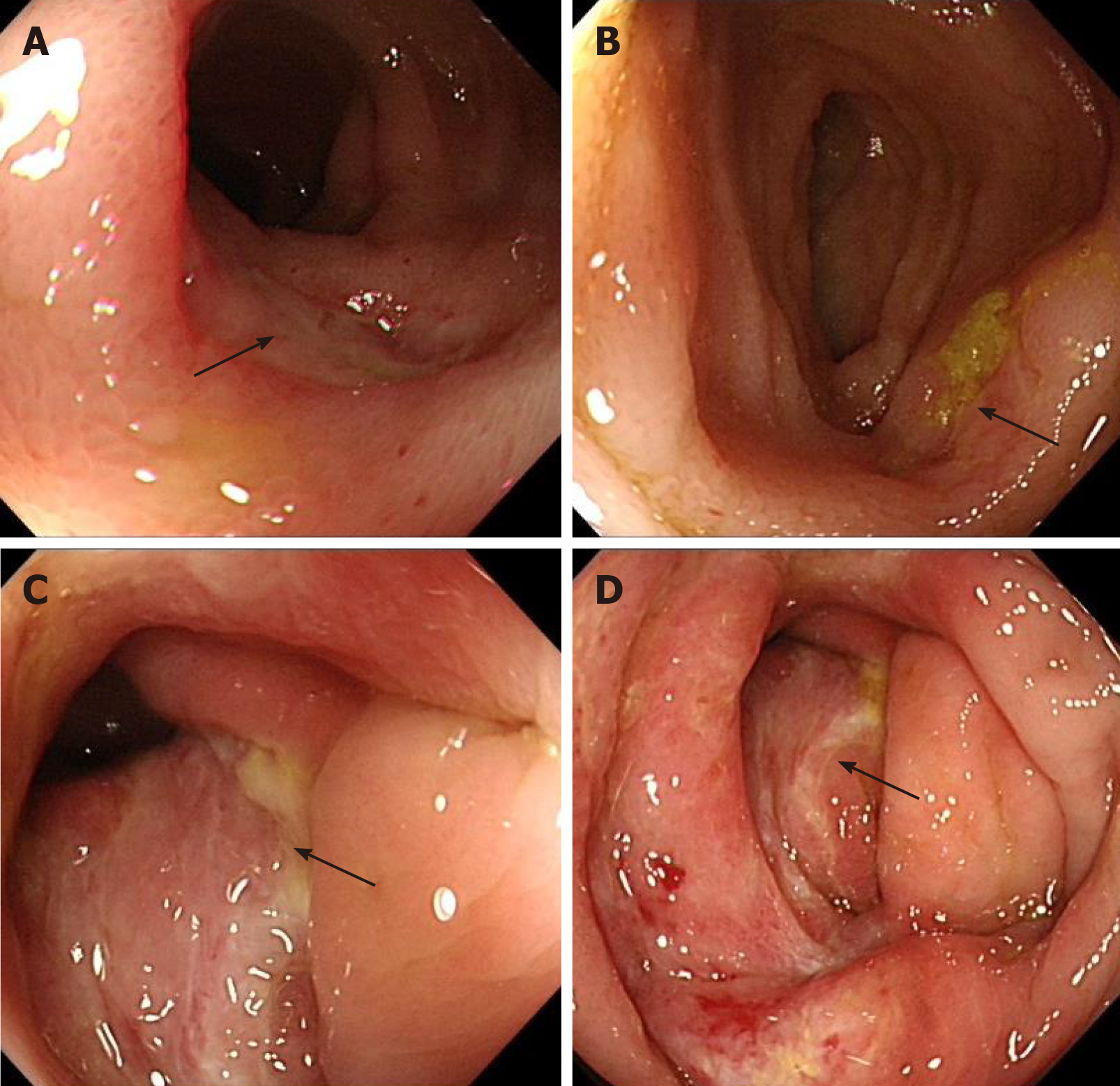
Figure 1 Colonoscopy before treatment.
There are multiple ulcers in the terminal ileum and the ileocecal region, as indicated by the black arrow. A: Ulcer in terminal ileum; B: Another ulcer in terminal ileum; C: Ulcer in ileocecal region; and D: Ulcer in ileocecal region.
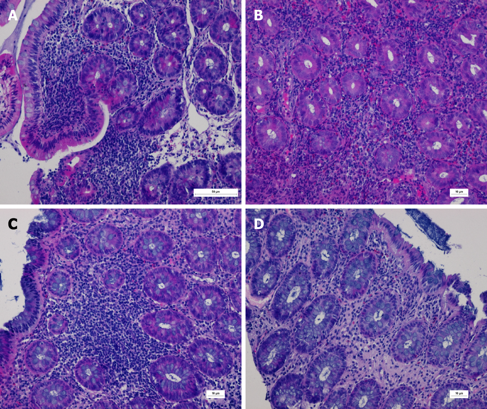
Figure 2 Prompted infiltration of diffuse inflammatory cells and scattered eosinophils, with visible ulcer formation.
Periodic acid-Schiff (-). Immunohistochemical cytomegalovirus (-). Epstein-Barr virus encoded ribonucleic acid (-). A: Terminal ileum, length of scale bar is 50 µm; B: Ileocecal region, length of scale bar is 10 µm; C: Descending colon, length of scale bar is 10 µm; D: Rectum, length of scale bar is 10 µm.
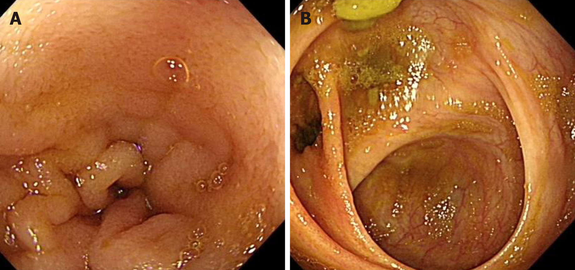
Figure 3 Colonoscopy after treatment.
Colonoscopy review after treatment for 6 mo. Mucosa in terminal ileum and ileocecal region become smooth. A: Terminal ileum; B: Ileocecal region.
- Citation: Gong YZ, Zhong XM, Zou JZ. Infliximab treatment of glycogenosis Ib with Crohn's-like enterocolitis: A case report. World J Clin Cases 2021; 9(19): 5280-5286
- URL: https://www.wjgnet.com/2307-8960/full/v9/i19/5280.htm
- DOI: https://dx.doi.org/10.12998/wjcc.v9.i19.5280


