Copyright
©The Author(s) 2021.
World J Clin Cases. Jul 6, 2021; 9(19): 5238-5244
Published online Jul 6, 2021. doi: 10.12998/wjcc.v9.i19.5238
Published online Jul 6, 2021. doi: 10.12998/wjcc.v9.i19.5238
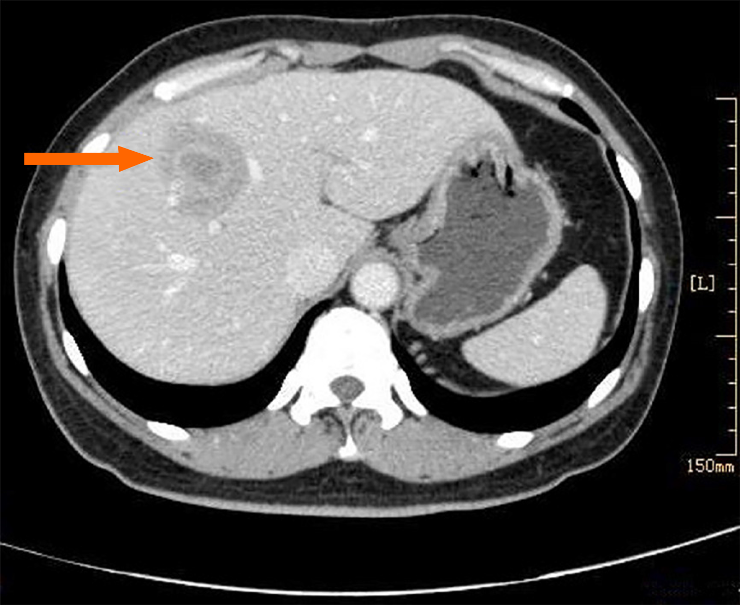
Figure 1 Enhancemed computed tomography revealed a low density mass shadow (orange arrow) with a distinct boundary in the anterior and superior segment of the right lobe of liver.
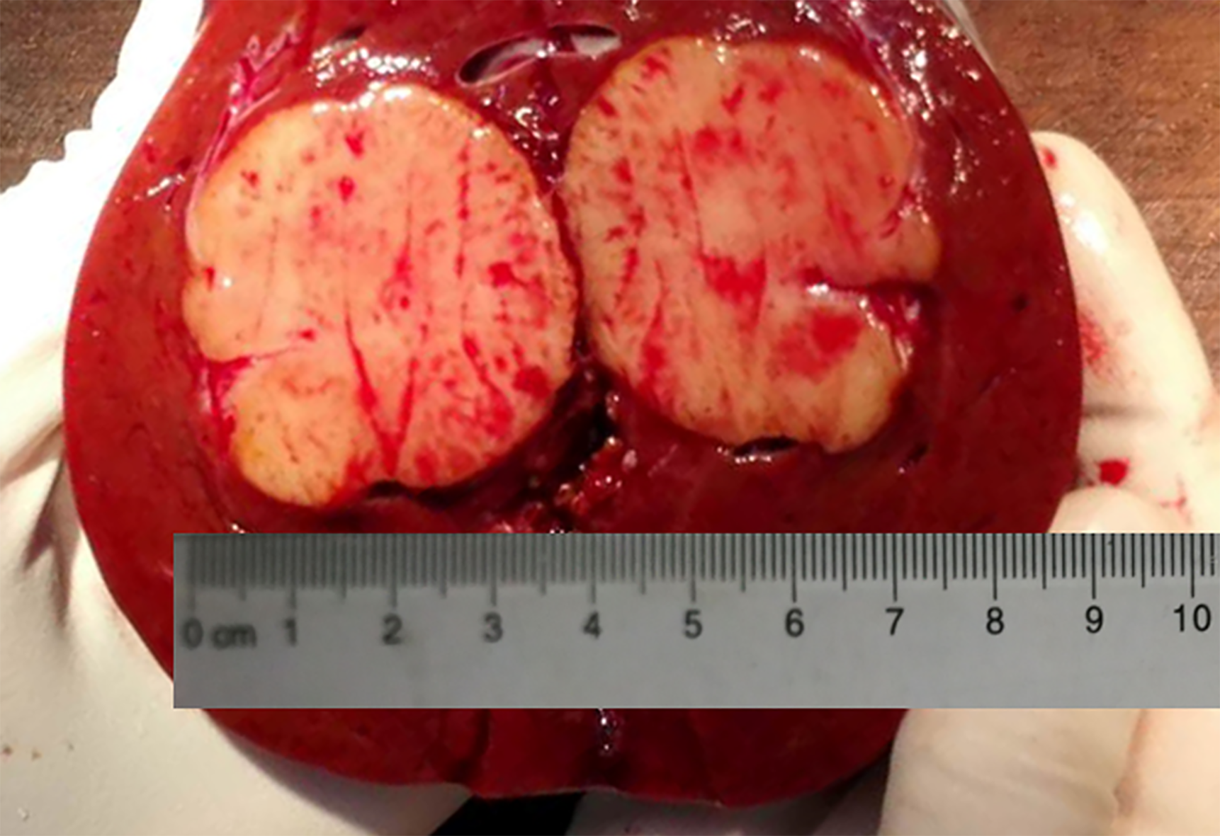
Figure 2 A 3.
8 cm × 3.5 cm × 3 cm grayish-white mass with distinct boundary located 1 cm under the capsule of the liver.
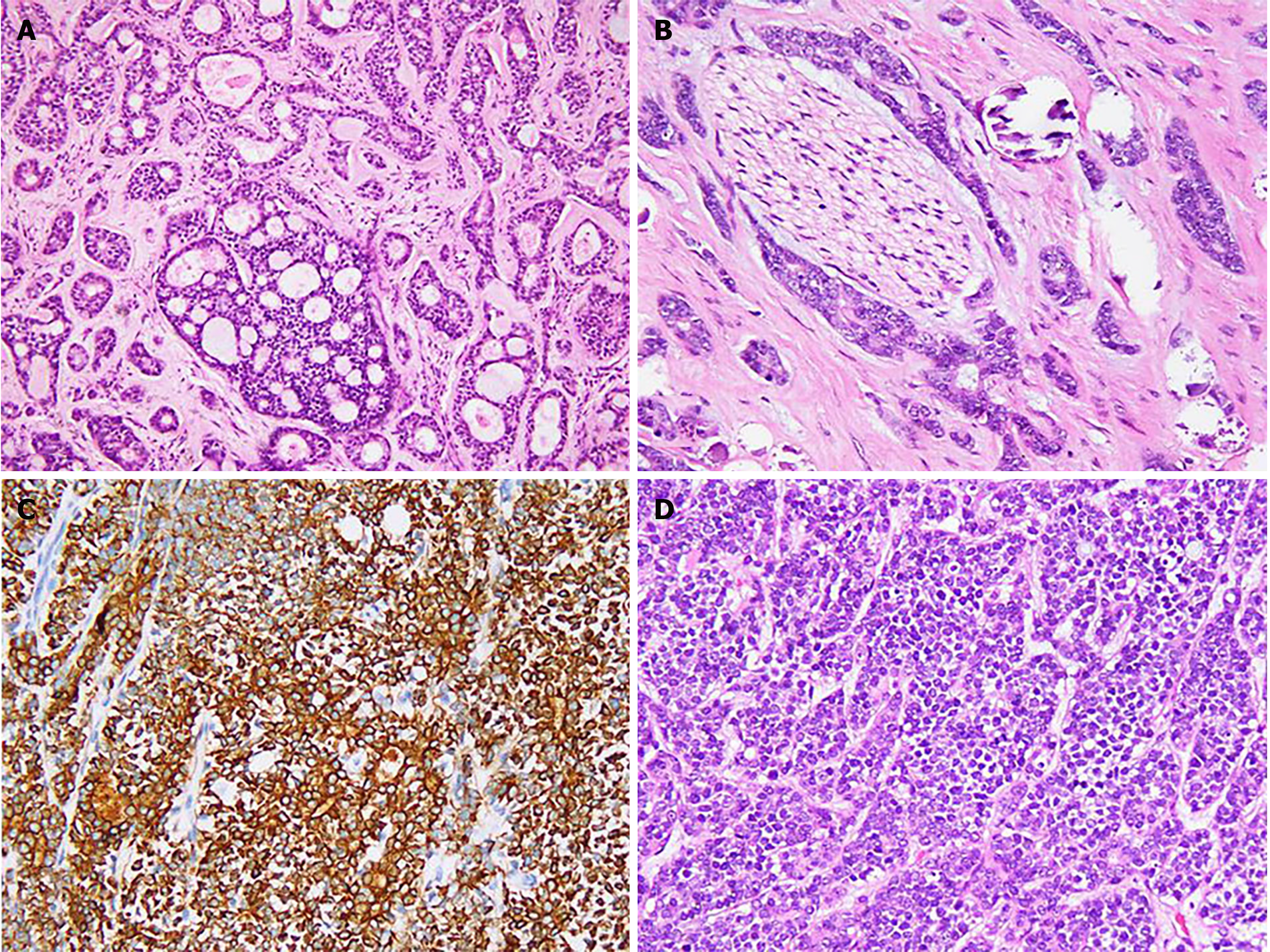
Figure 3 Histopathological findings of adenoid cystic carcinoma.
A: Tubiform and cribriform architecture of metastatic adenoid cystic carcinoma (ACC) of the liver (hematoxylin-eosin, × 200); B: ACC involvement of nerve, × 200); C: Immunostaining showed ductal cells positive for CD117 (DAB, × 200); D: Solid architecture in the sublingual gland ACC (hematoxylin-eosin, × 200).
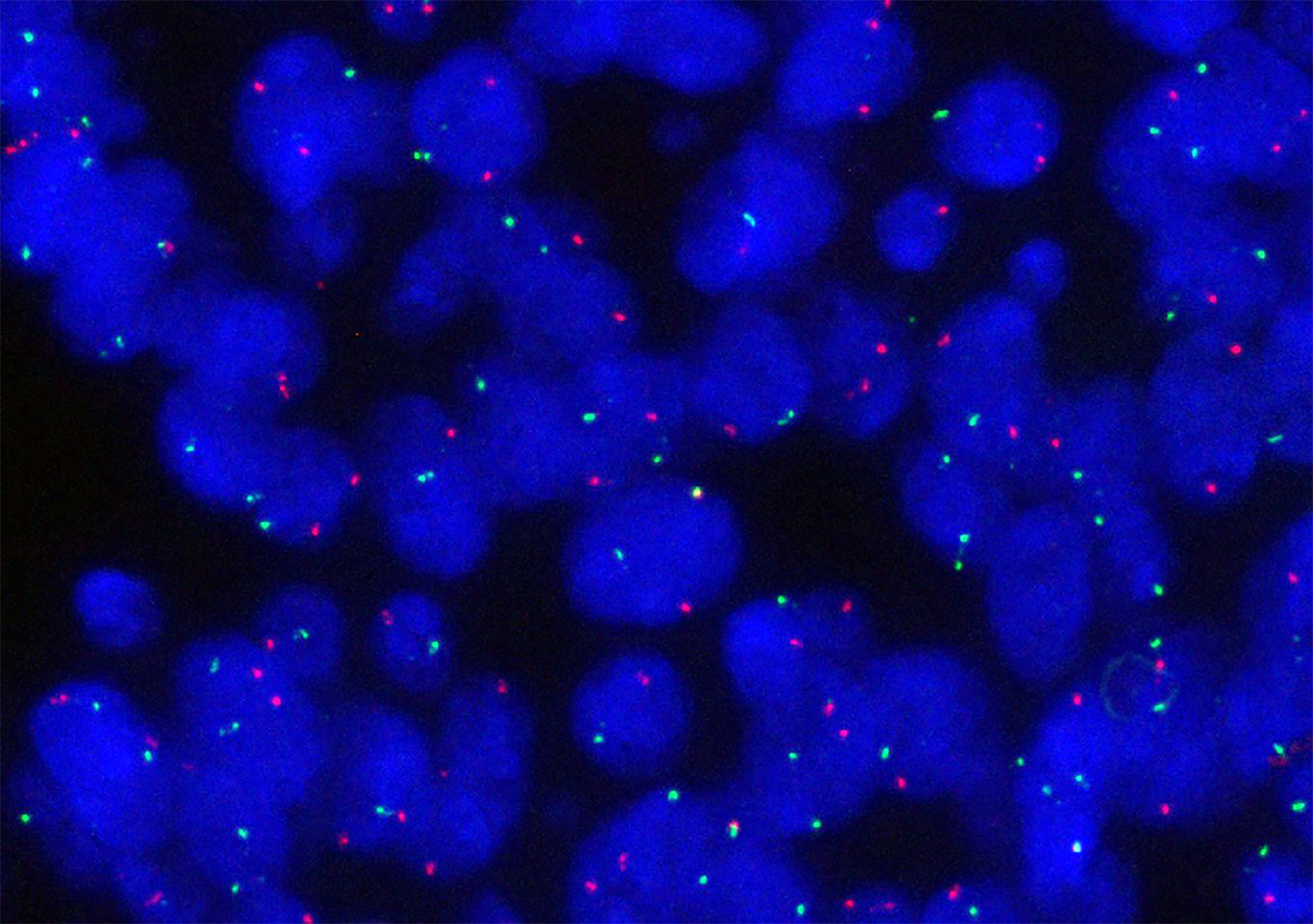
Figure 4 MYB-NFIB fusion gene was not detected by fluorescence in situ hybridization in either the metastatic adenoid cystic carcinoma of liver or the primary adenoid cystic carcinoma of the sublingual gland.
- Citation: Li XH, Zhang YT, Feng H. Liver metastasis as the initial clinical manifestation of sublingual gland adenoid cystic carcinoma: A case report. World J Clin Cases 2021; 9(19): 5238-5244
- URL: https://www.wjgnet.com/2307-8960/full/v9/i19/5238.htm
- DOI: https://dx.doi.org/10.12998/wjcc.v9.i19.5238


