Copyright
©The Author(s) 2021.
World J Clin Cases. Jul 6, 2021; 9(19): 5232-5237
Published online Jul 6, 2021. doi: 10.12998/wjcc.v9.i19.5232
Published online Jul 6, 2021. doi: 10.12998/wjcc.v9.i19.5232
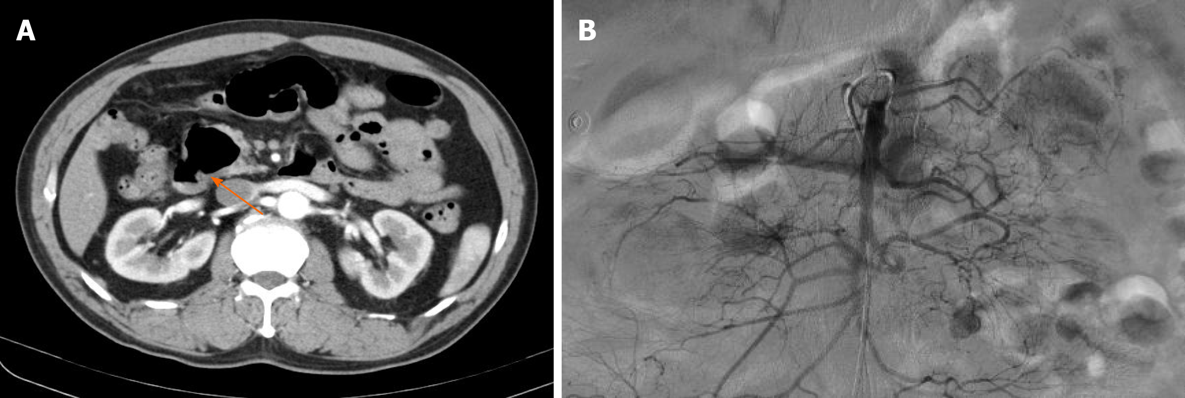
Figure 1 Preoperative image examinations.
A: The abdominal computed tomography showed dilated small bowel loops with multiple jejunal diverticula (orange arrow); B: No visible sites of bleeding were shown according to the mesenteric angiography.
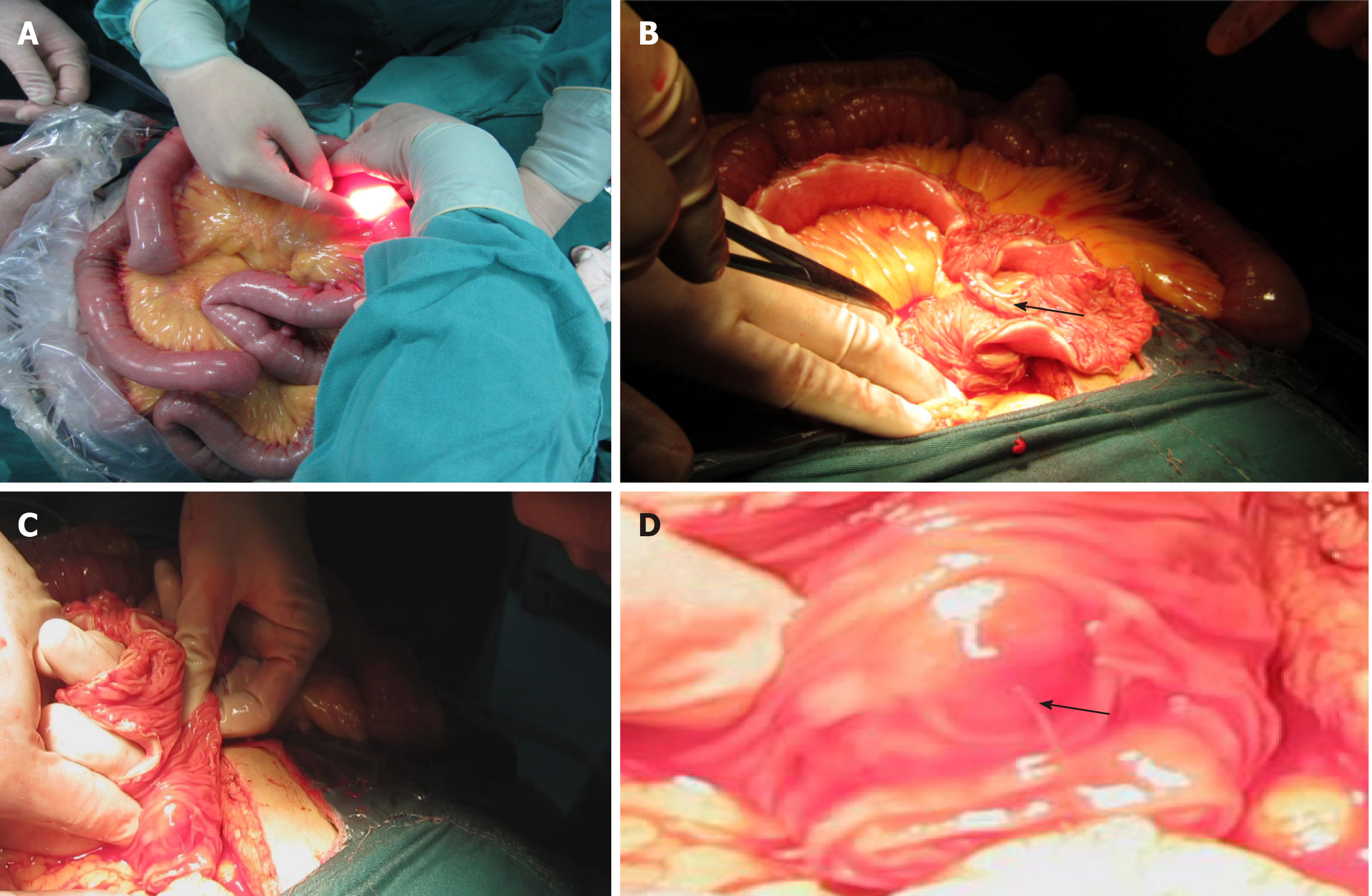
Figure 2 Intraoperative images findings.
A: The gastroscope was inserted into the lumen via a small incision in the jejunum; B: The full-thickness enterotomy with exploration was performed (black arrow), and the diverticula were detected from proximal to distal under direct vision; C: A pulsating vessel was identified in the first diverticulum under the duodeno-jejunal flexure; D: The magnified rectangle in Figure 2C, the actively bleeding clearly shown (black arrow).
- Citation: Ma HC, Xiao H, Qu H, Wang ZJ. Successful diagnosis and treatment of jejunal diverticular haemorrhage by full-thickness enterotomy: A case report. World J Clin Cases 2021; 9(19): 5232-5237
- URL: https://www.wjgnet.com/2307-8960/full/v9/i19/5232.htm
- DOI: https://dx.doi.org/10.12998/wjcc.v9.i19.5232


