Copyright
©The Author(s) 2021.
World J Clin Cases. Jul 6, 2021; 9(19): 5217-5225
Published online Jul 6, 2021. doi: 10.12998/wjcc.v9.i19.5217
Published online Jul 6, 2021. doi: 10.12998/wjcc.v9.i19.5217
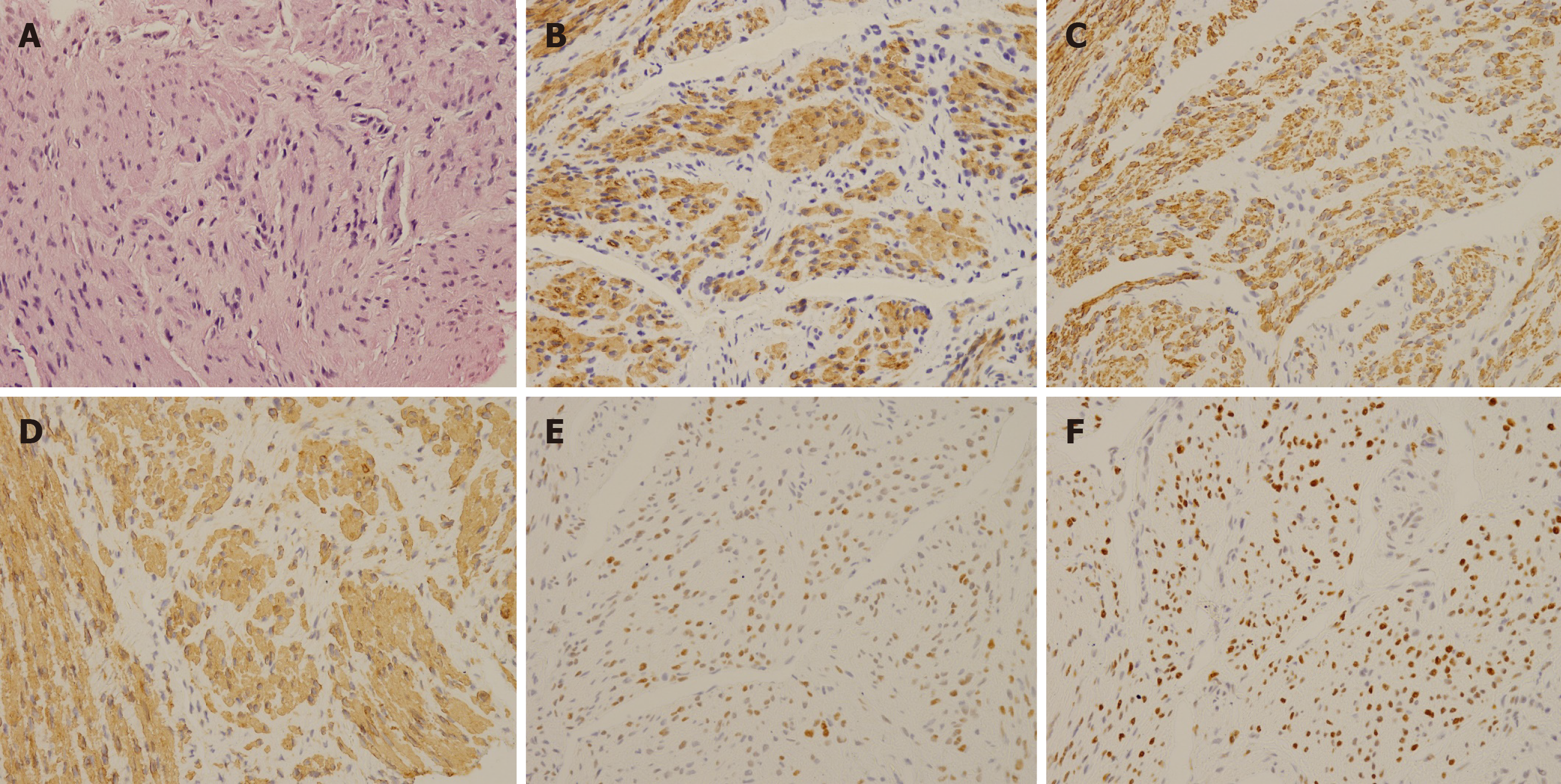
Figure 1 Pathological examination of leiomyomatosis peritonealis disseminate.
A: Hematoxylin-eosin staining of tumor cells showed a spindle-shaped smooth-muscle cell tumor without necrosis and atypia (400-fold); B-F: Immunohistochemical staining of smooth-muscle cells showed that Caldesmon (B), Desmin (C), smooth-muscle actin (D), estrogen receptors (E), and progesterone receptors (F) were positive (400-fold).
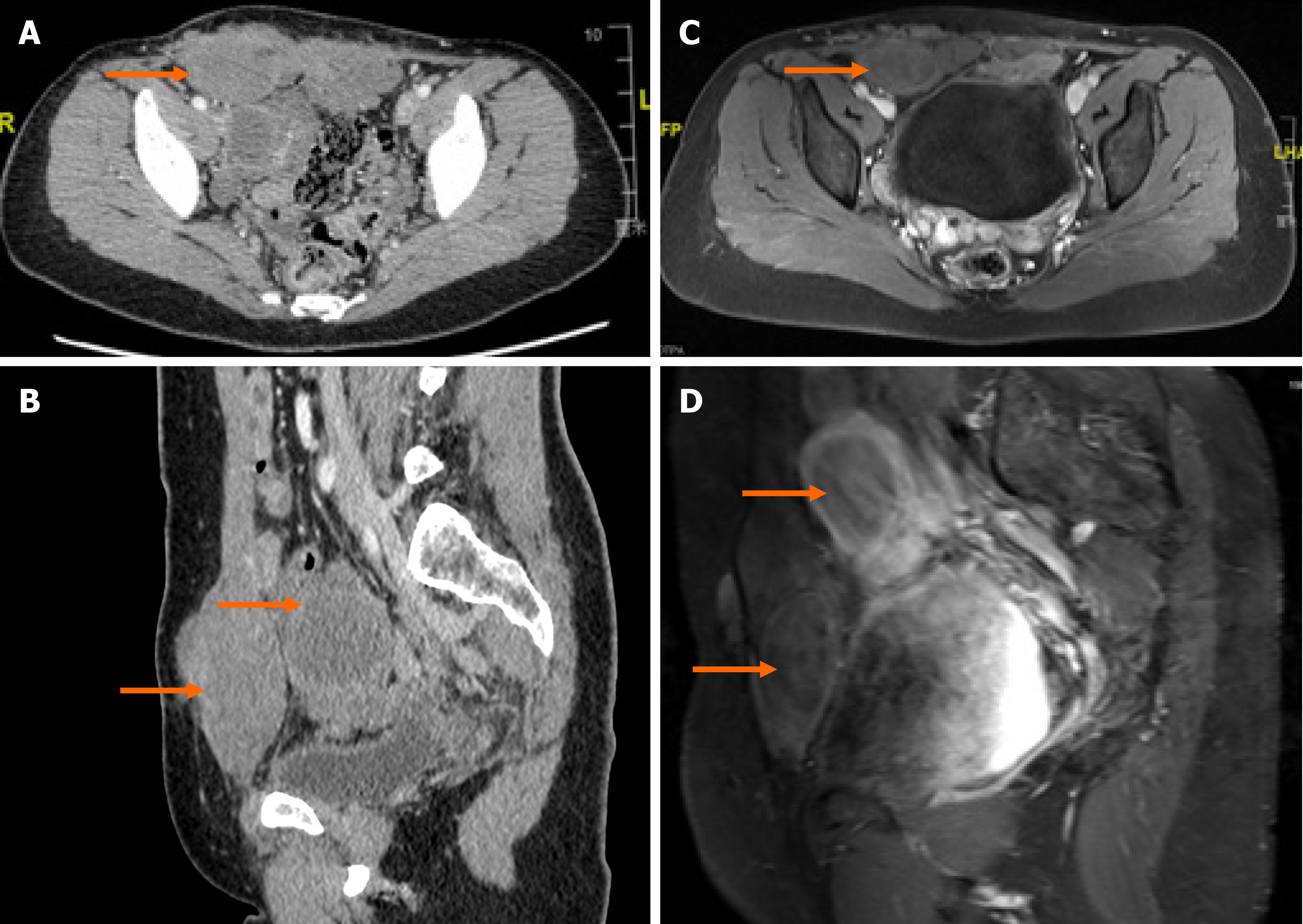
Figure 2 Computed tomography and magnetic resonance imaging scans before and after treatment.
The red arrows indicate the nodules involving the abdominal wall and pelvic cavity. A: Computed tomography (CT) scan of the pelvis in the transverse plane in January 2018; B: CT scan of the pelvis in the sagittal plane in January 2018; C: Magnetic resonance imaging (MRI) scan of the pelvis in the transverse plane in December 2019; D: MRI scan of the pelvis in the sagittal plane in December 2019.
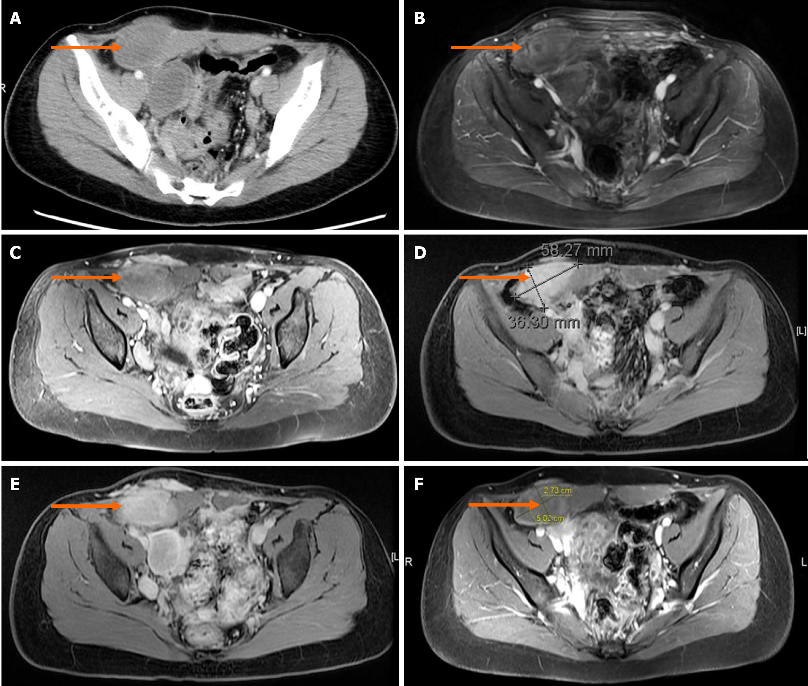
Figure 3 Comparison before and after treatment.
A: Computed tomography scan of the biggest nodule before treatment in January 2018; B: Magnetic resonance imaging (MRI) scan of the biggest nodule after tamoxifen therapy in July 2018; C: MRI scan of the biggest nodule after first goserelin acetate therapy in January 2019; D: MRI scan of the biggest nodule after ulipristal acetate treatment in June 2019; E: MRI scan of the biggest nodule after second goserelin acetate therapy in December 2019; F: MRI scan of the biggest nodule in October 2020.
- Citation: Yang JW, Hua Y, Xu H, He L, Huo HZ, Zhu CF. Treatment of leiomyomatosis peritonealis disseminata with goserelin acetate: A case report and review of the literature. World J Clin Cases 2021; 9(19): 5217-5225
- URL: https://www.wjgnet.com/2307-8960/full/v9/i19/5217.htm
- DOI: https://dx.doi.org/10.12998/wjcc.v9.i19.5217


