Copyright
©The Author(s) 2021.
World J Clin Cases. Jul 6, 2021; 9(19): 5197-5202
Published online Jul 6, 2021. doi: 10.12998/wjcc.v9.i19.5197
Published online Jul 6, 2021. doi: 10.12998/wjcc.v9.i19.5197
Figure 1 Imaging findings in a 40-year-old woman with peliosis hepatis.
A: Plain computed tomography image showed multiple high- and low-density stratification signs (white arrows) within a massive cystic lesion in the right lobe; B: Enhanced computed tomography scan image showed slight enhancement in the margin of the lesion with density lower than that of the surrounding liver parenchyma (white arrow); C: Plain magnetic resonance imaging scan T2WI image showed massive “capsule-in-capsule”-like lesions in the liver parenchyma, mixed lesion signals, primarily high signal. Fluid–fluid (high-low signal) levels (white arrows) were visible in the capsule.
Figure 2 Postoperative pathology.
Hematoxylin and eosin staining, 100× (A) and hematoxylin and eosin staining, 200× (B) microscopy showed multiple blood-filled cysts and liver parenchymal hemorrhage and necrosis in the lesion. The inner wall of the cyst cavity was not lined by endothelial cells. These results were consistent with the presentation of peliosis hepatis.
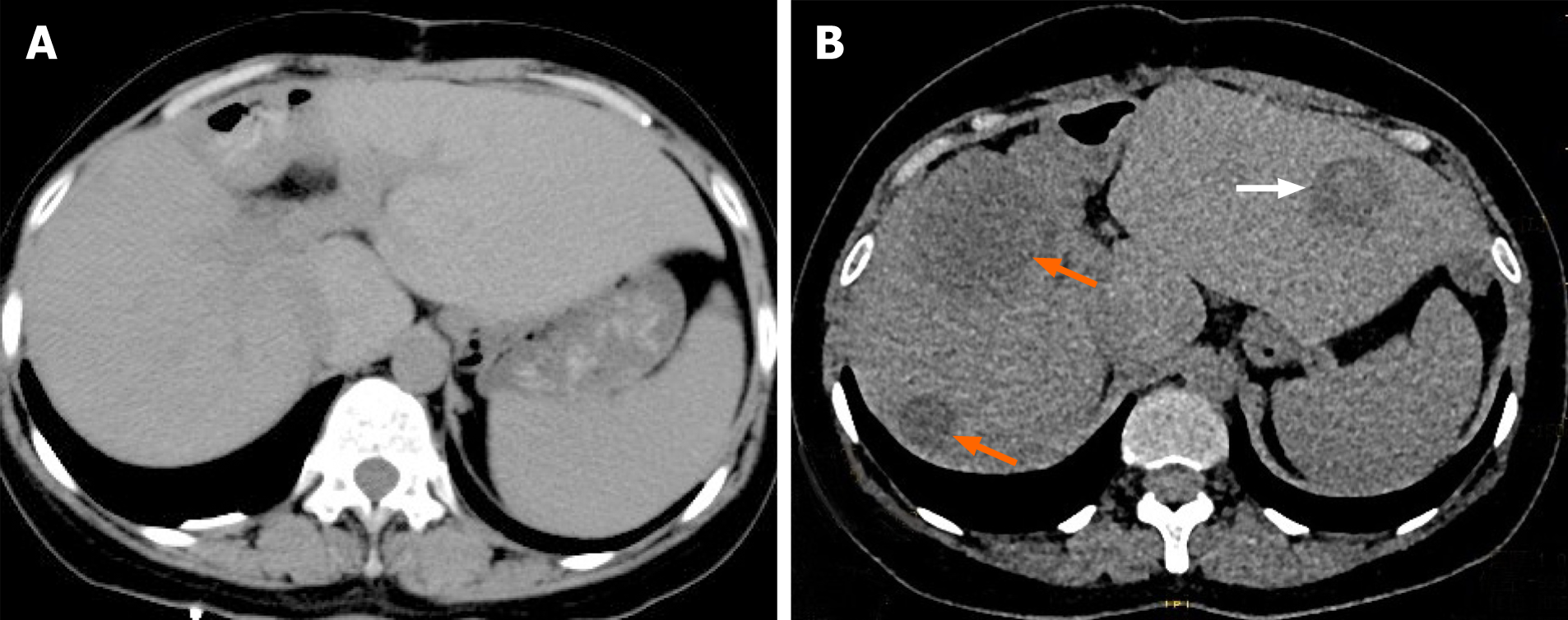
Figure 3 Postoperative re-examination images.
A: Computed tomography re-examination at 3 mo postoperatively did not indicate obvious abnormal density opacities in the liver parenchyma; B: Computed tomography re-examination at 1 year postoperatively revealed multiple patches of low- or slightly low-density opacities in the liver parenchyma (orange arrow), with patchy high-density hemorrhage in the lesion (white arrow).
- Citation: Ren SX, Li PP, Shi HP, Chen JH, Deng ZP, Zhang XE. Imaging presentation and postoperative recurrence of peliosis hepatis: A case report. World J Clin Cases 2021; 9(19): 5197-5202
- URL: https://www.wjgnet.com/2307-8960/full/v9/i19/5197.htm
- DOI: https://dx.doi.org/10.12998/wjcc.v9.i19.5197


