Copyright
©The Author(s) 2021.
World J Clin Cases. Jul 6, 2021; 9(19): 4969-4979
Published online Jul 6, 2021. doi: 10.12998/wjcc.v9.i19.4969
Published online Jul 6, 2021. doi: 10.12998/wjcc.v9.i19.4969
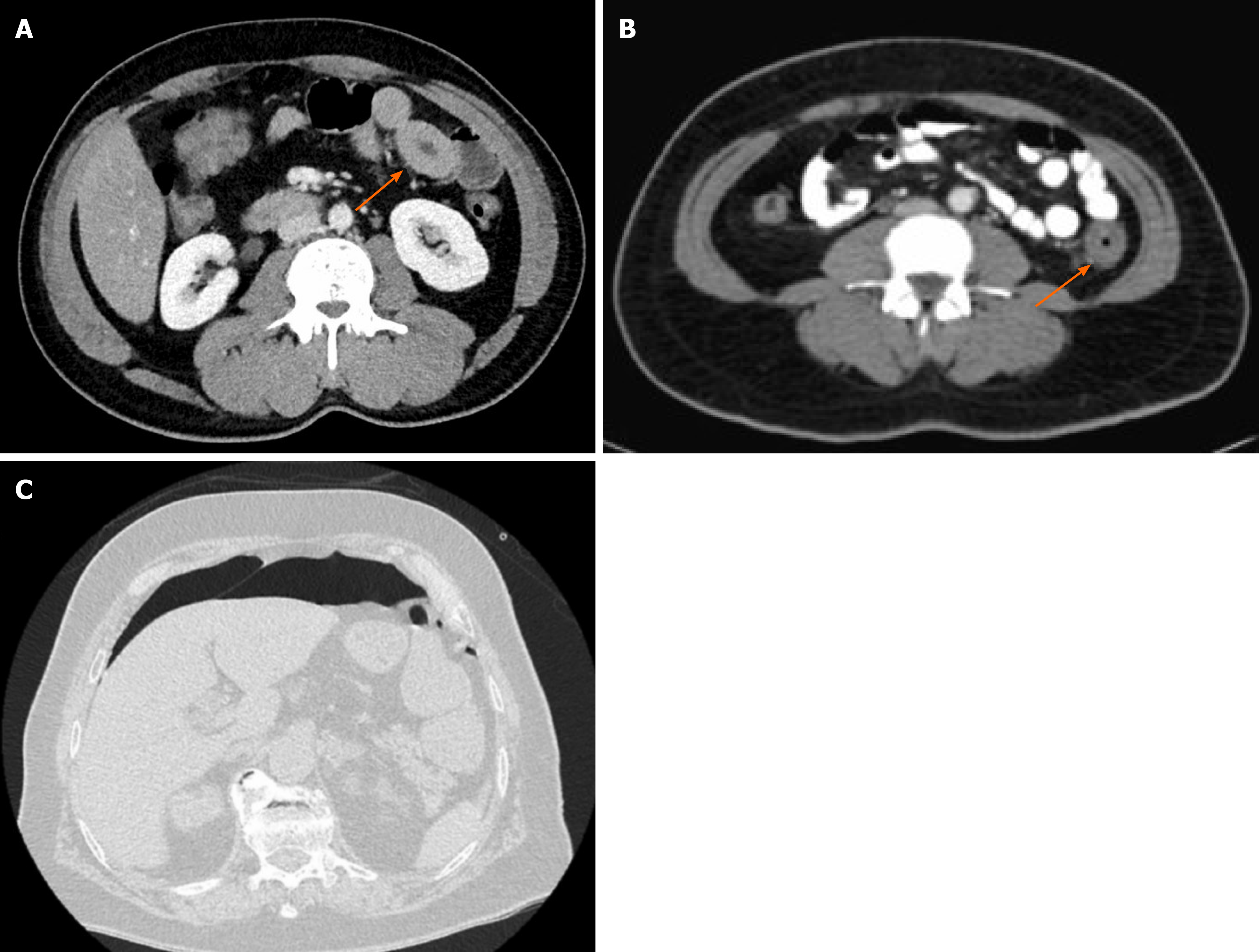
Figure 1 Computed tomography images[22,23].
A: Thick-walled loop of small bowel (arrow) with mild perienteric fat stranding in computed tomography (CT) abdominal image; B: Thick-walled loop of descending colon (arrow) with mild perienteric fat stranding in CT image; C: Extensive pneumoperitoneum caused by perforation of the sigmoid colon on abdominal CT image.
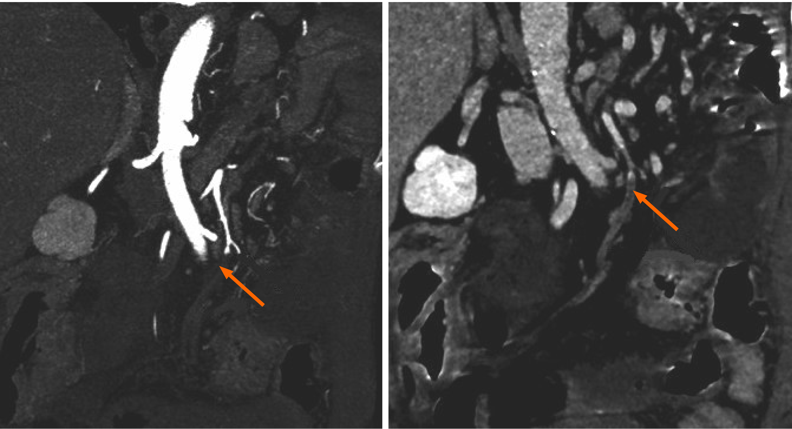
Figure 2 Thrombosis of the superior mesenteric artery in a severe acute respiratory syndrome coronavirus 2 infected patient’s computed tomography images[28].
Arrows: Thrombosis of the superior mesenteric artery.
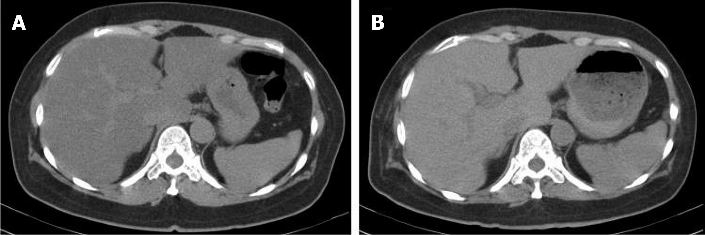
Figure 3 An inpatient coronavirus disease 2019 patient’s abdominal computed tomography image[55].
A: Decreased hepatic computed tomography (CT) attenuation value and liver-to-spleen attenuation (L/S) ratio; B The hepatic CT attenuation value and L/S ratio returned to normal level after intensive therapies.
- Citation: Fang LG, Zhou Q. Remarkable gastrointestinal and liver manifestations of COVID-19: A clinical and radiologic overview. World J Clin Cases 2021; 9(19): 4969-4979
- URL: https://www.wjgnet.com/2307-8960/full/v9/i19/4969.htm
- DOI: https://dx.doi.org/10.12998/wjcc.v9.i19.4969


