Copyright
©The Author(s) 2021.
World J Clin Cases. Jun 26, 2021; 9(18): 4866-4872
Published online Jun 26, 2021. doi: 10.12998/wjcc.v9.i18.4866
Published online Jun 26, 2021. doi: 10.12998/wjcc.v9.i18.4866
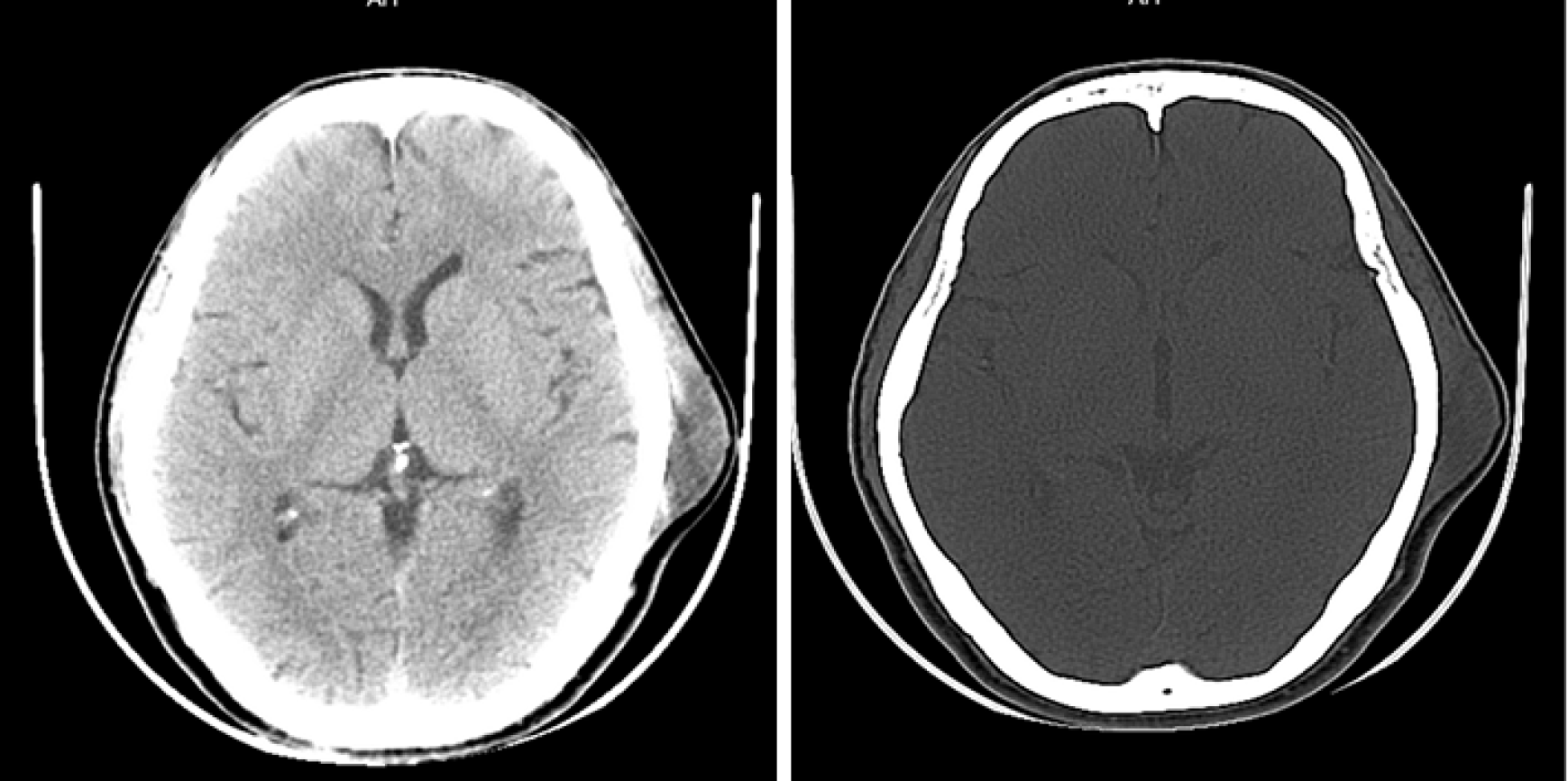
Figure 1 Computed tomography shows a subcutaneous mass in the left temporal region without skull destruction.
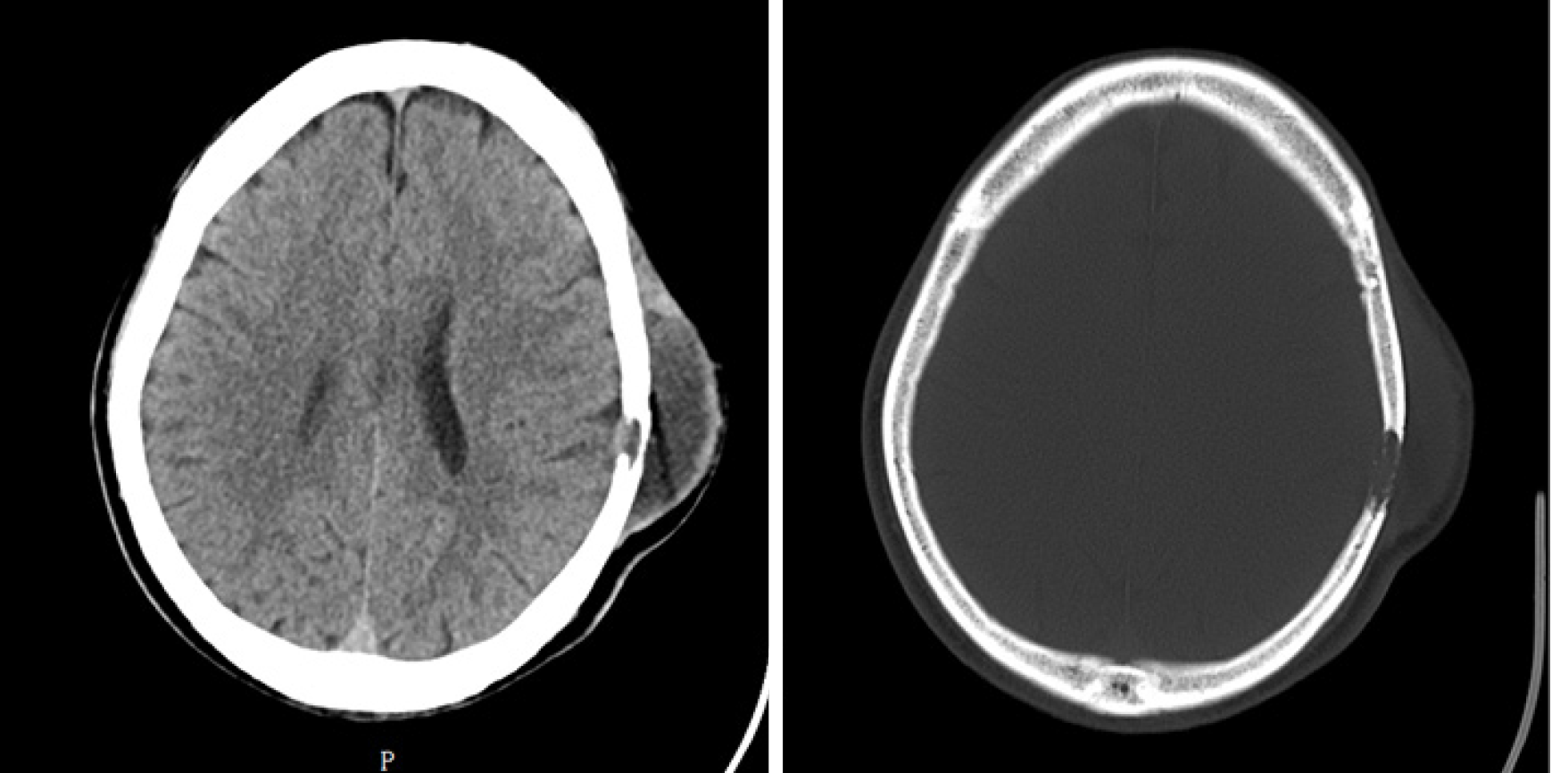
Figure 2 Computed tomography shows a soft tissue mass in the left temporal region and destruction of the adjacent skull.
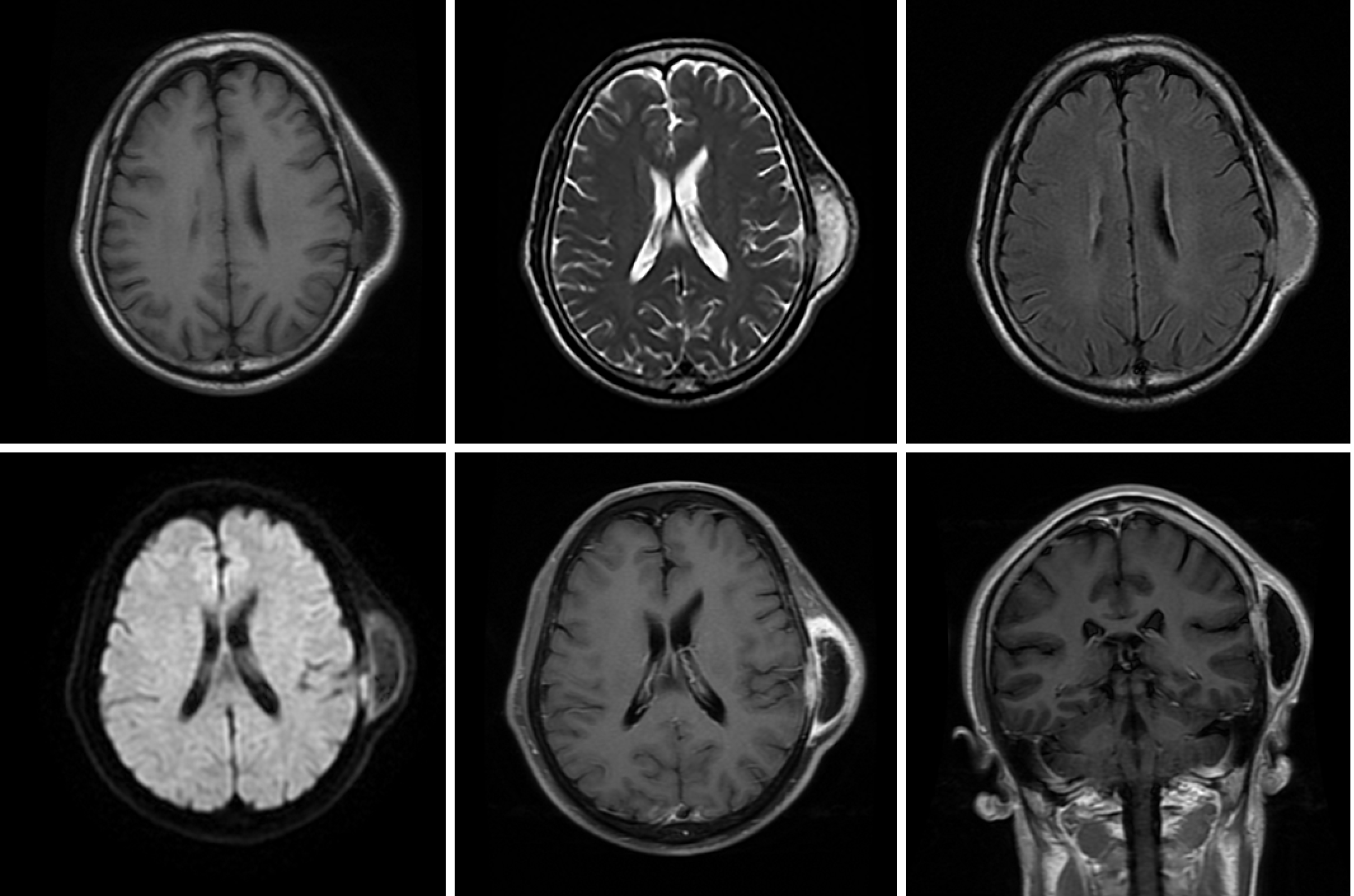
Figure 3 Magnetic resonance imaging shows a mass in the left temporal muscle, with obvious enhancement around the tumor, but no enhancement in the center of the tumor.
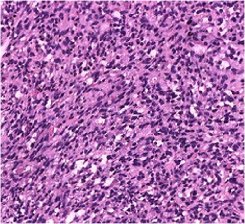
Figure 4 Pathological examination revealed a tumor composed of mildly atypical spindle cells arranged in a crossed bundle or spiral, and with a few rhabdomyoblasts scattered among the spindle cells (hematoxylin and eosin, × 200).
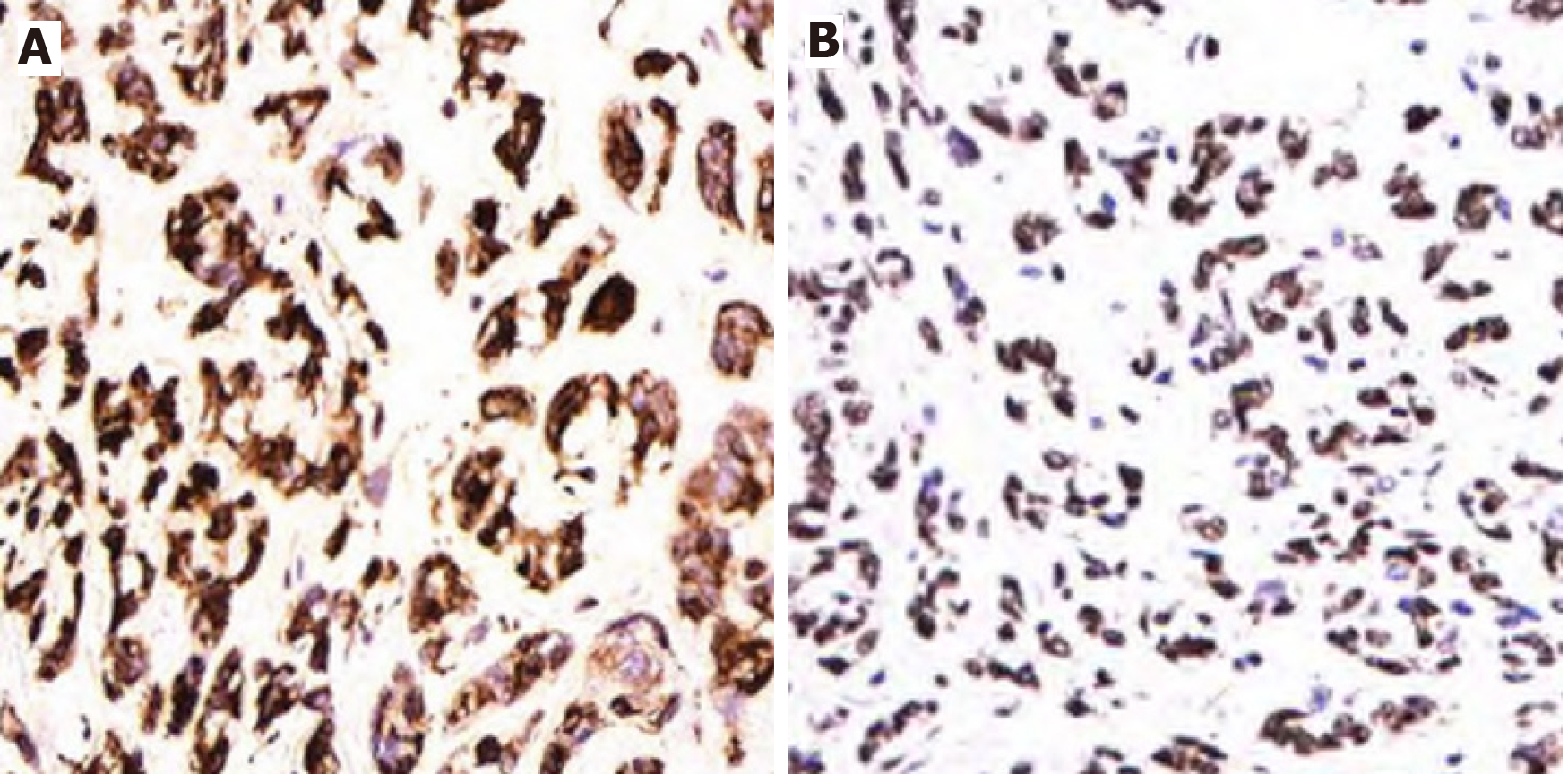
Figure 5 Immunohistochemical staining of tumor tissue.
A: Desmin is strongly positive in the cytoplasm of tumor cells (× 200); B: MyoD1 is strongly positive in the nucleus of tumor cells (× 200).
- Citation: Wang GH, Shen HP, Chu ZM, Shen J. Adult rhabdomyosarcoma originating in the temporal muscle, invading the skull and meninges: A case report. World J Clin Cases 2021; 9(18): 4866-4872
- URL: https://www.wjgnet.com/2307-8960/full/v9/i18/4866.htm
- DOI: https://dx.doi.org/10.12998/wjcc.v9.i18.4866


