Copyright
©The Author(s) 2021.
World J Clin Cases. Jun 26, 2021; 9(18): 4859-4865
Published online Jun 26, 2021. doi: 10.12998/wjcc.v9.i18.4859
Published online Jun 26, 2021. doi: 10.12998/wjcc.v9.i18.4859
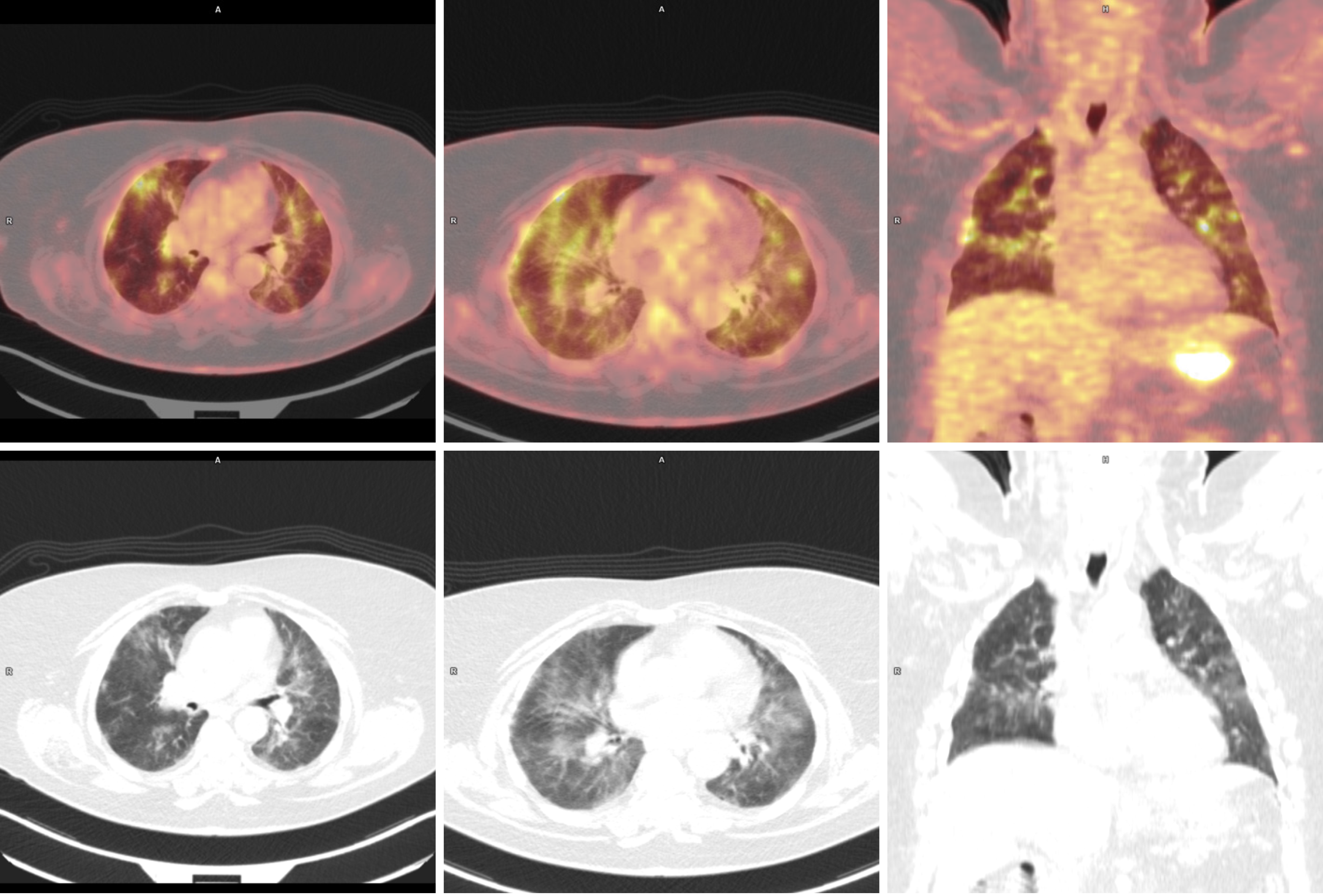
Figure 1 Radiological findings from positron emission tomography-computed tomography.
Regression in metabolism and measurement of affected lymph nodes and spleen. Previously nonexistent pulmonary changes, metabolically active, disseminated areas of pulmonary parenchyma in both lungs, with central and subpleural distribution, with greater intensity in the upper and middle lung areas.
- Citation: Łącki S, Wyżgolik K, Nicze M, Georgiew-Nadziakiewicz S, Chudek J, Wdowiak K. Low symptomatic COVID-19 in an elderly patient with follicular lymphoma treated with rituximab-based immunotherapy: A case report. World J Clin Cases 2021; 9(18): 4859-4865
- URL: https://www.wjgnet.com/2307-8960/full/v9/i18/4859.htm
- DOI: https://dx.doi.org/10.12998/wjcc.v9.i18.4859


