Copyright
©The Author(s) 2021.
World J Clin Cases. Jun 26, 2021; 9(18): 4829-4836
Published online Jun 26, 2021. doi: 10.12998/wjcc.v9.i18.4829
Published online Jun 26, 2021. doi: 10.12998/wjcc.v9.i18.4829
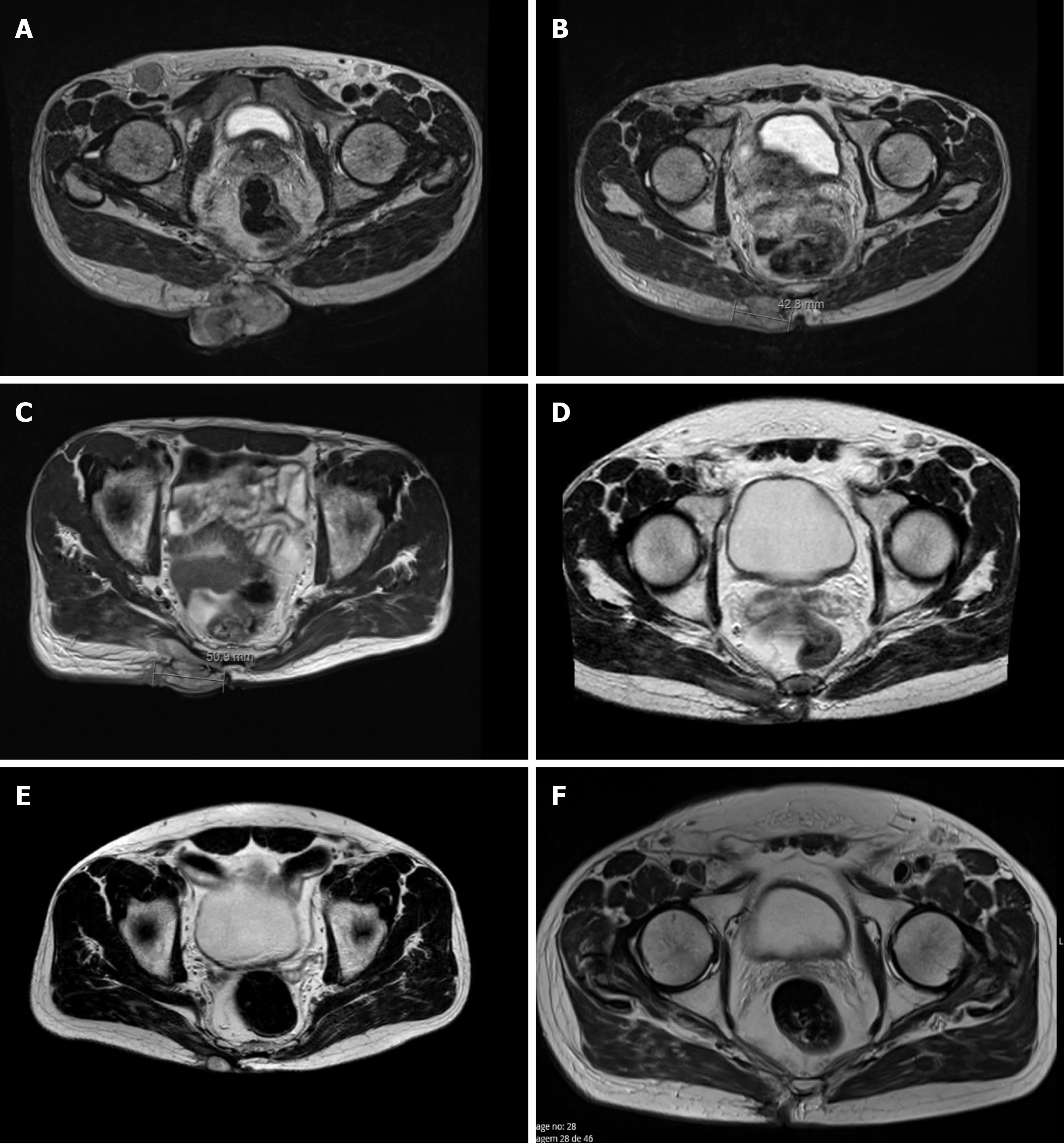
Figure 1 Magnetic resonance imaging images of the primary Merkel cell carcinoma lesion.
A: Baseline; B: After two cycles of chemotherapy (partial response); C: After six cycles of chemotherapy (progression of the disease); D: After seven cycles of avelumab (partial response); E: After 14 cycles of avelumab (partial response); F: After 28 cycles of avelumab.
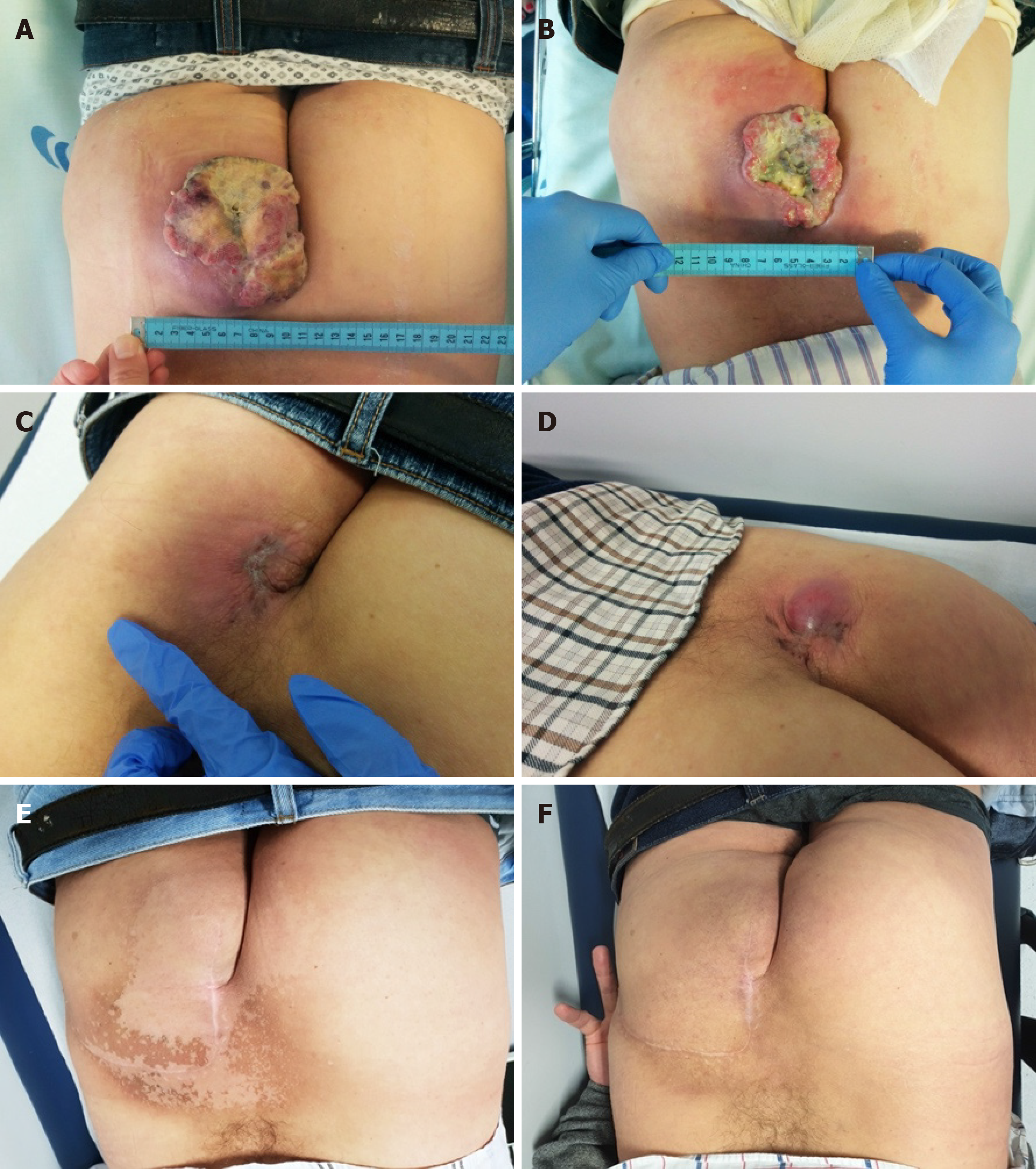
Figure 2 Photographic registries of the evolution of the primary Merkel cell carcinoma lesion.
A: Baseline avelumab; B: After two cycles of avelumab; C: After seven cycles of avelumab; D: After 16 cycles of avelumab; E: After surgery, radiotherapy, and 28 cycles of avelumab; F: Follow-up, more than 2.5 years after surgery.
- Citation: Leão I, Marinho J, Costa T. Long-term response to avelumab and management of oligoprogression in Merkel cell carcinoma: A case report. World J Clin Cases 2021; 9(18): 4829-4836
- URL: https://www.wjgnet.com/2307-8960/full/v9/i18/4829.htm
- DOI: https://dx.doi.org/10.12998/wjcc.v9.i18.4829


