Copyright
©The Author(s) 2021.
World J Clin Cases. Jun 26, 2021; 9(18): 4783-4788
Published online Jun 26, 2021. doi: 10.12998/wjcc.v9.i18.4783
Published online Jun 26, 2021. doi: 10.12998/wjcc.v9.i18.4783
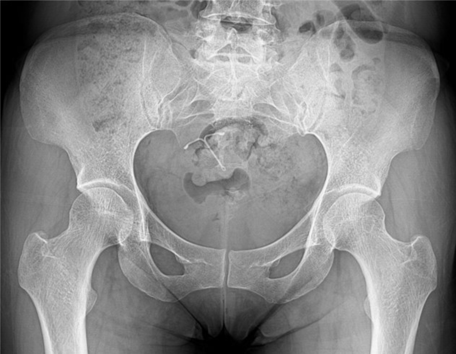
Figure 1 Simple radiography of the hip joint: Herniation pit, a small thin sclerotic rimmed radiolucent lesion of the left femoral head.
Figure 2 Hip magnetic resonance imaging showed signal elevation of both femur necks.
A: T1 image; B: T2 image.
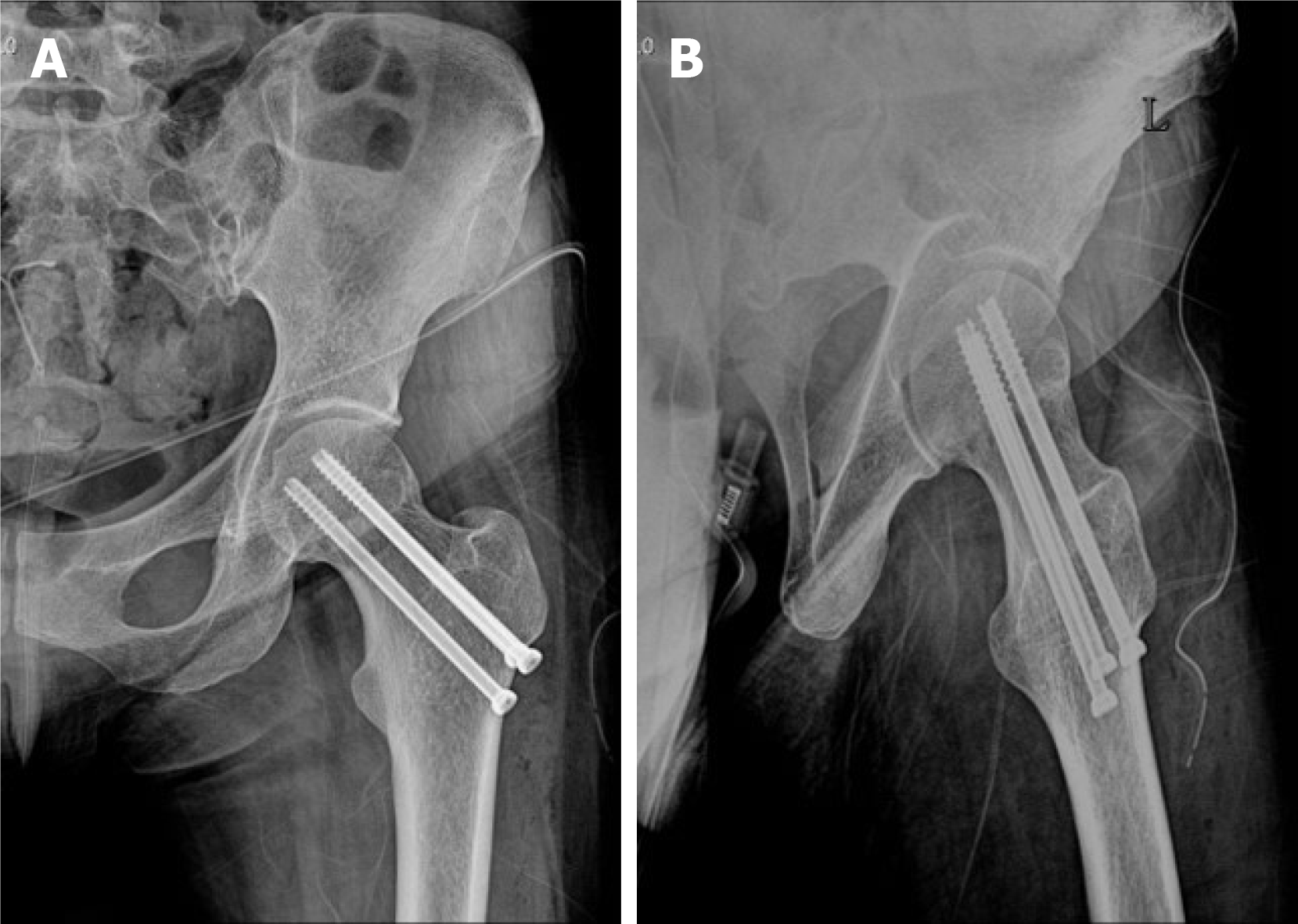
Figure 3 Simple radiography of closed reduction and internal fixation using a cannulated screw at postoperative 2 mo.
A: Antero-posterior image; B: Lateral image.
Figure 4 Follow-up magnetic resonance imaging findings.
A: T1 image; B: T2 image.
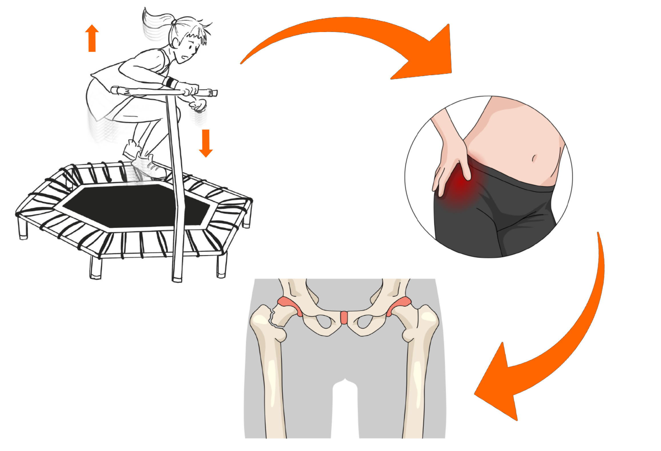
Figure 5 Schematic animation of stress fracture of the femoral neck after trampoline exercise.
- Citation: Nam DC, Hwang SC, Lee EC, Song MG, Yoo JI. Femoral neck stress fractures after trampoline exercise: A case report . World J Clin Cases 2021; 9(18): 4783-4788
- URL: https://www.wjgnet.com/2307-8960/full/v9/i18/4783.htm
- DOI: https://dx.doi.org/10.12998/wjcc.v9.i18.4783


