Copyright
©The Author(s) 2021.
World J Clin Cases. Jun 26, 2021; 9(18): 4778-4782
Published online Jun 26, 2021. doi: 10.12998/wjcc.v9.i18.4778
Published online Jun 26, 2021. doi: 10.12998/wjcc.v9.i18.4778
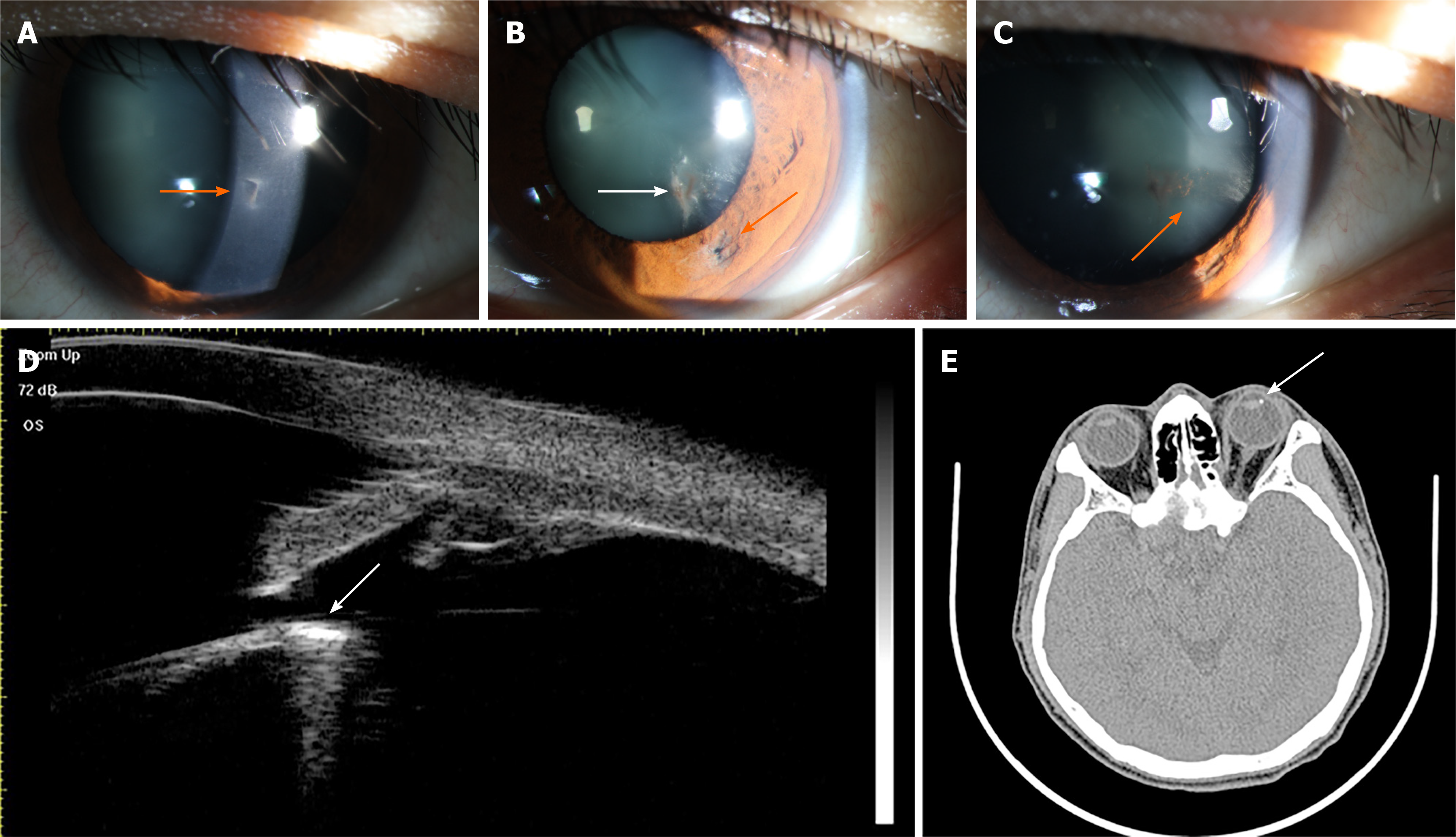
Figure 1 Examination of the injured eye preoperatively.
A-C: Anterior segment photographs. Panel A shows a well-closed full-thickness corneal perforation wound (orange arrow), panel B shows an iris hole (orange arrow) and only a localized lens opacity (white arrow) at the corresponding positions, and panel C shows a band of a cloudy area that was seen instead extending toward the equator of the lens (orange arrow); D: Computed tomography showed the relative positions of the foreign body and the lens (white arrow); E: Ultrasound biomicroscope examination confirmed the presence of the foreign body around the equator of the lens (white arrow).
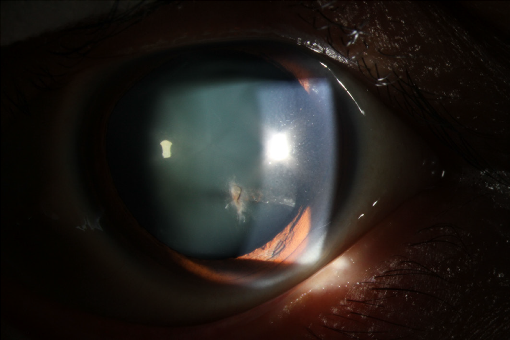
Figure 2 Anterior segment photograph of the injured eye 1 wk after surgery.
- Citation: Xue C, Chen Y, Gao YL, Zhang N, Wang Y. Disappeared intralenticular foreign body: A case report. World J Clin Cases 2021; 9(18): 4778-4782
- URL: https://www.wjgnet.com/2307-8960/full/v9/i18/4778.htm
- DOI: https://dx.doi.org/10.12998/wjcc.v9.i18.4778


