Copyright
©The Author(s) 2021.
World J Clin Cases. Jun 26, 2021; 9(18): 4760-4764
Published online Jun 26, 2021. doi: 10.12998/wjcc.v9.i18.4760
Published online Jun 26, 2021. doi: 10.12998/wjcc.v9.i18.4760
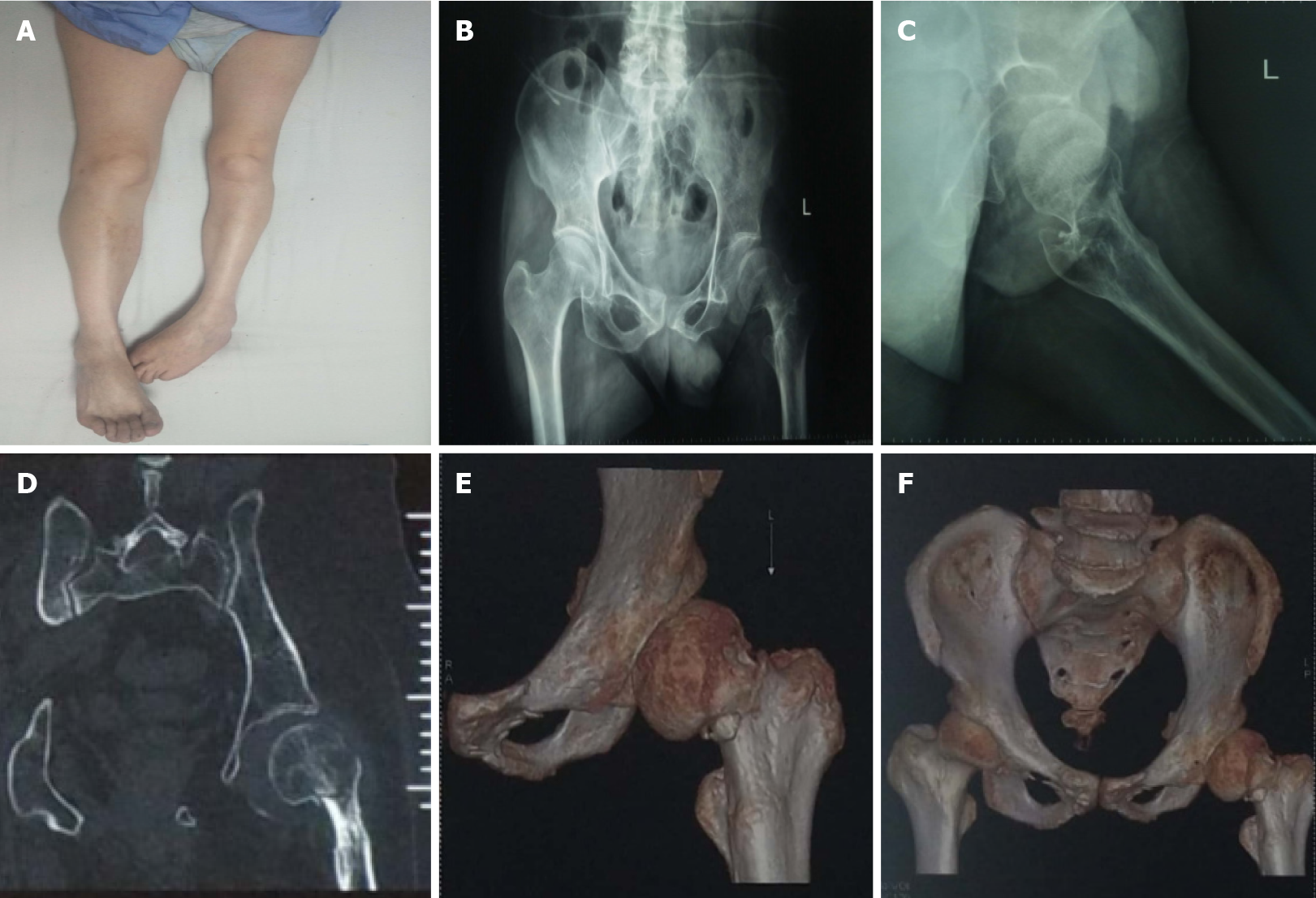
Figure 1 Preoperative data.
A: Preoperative general picture of both lower limbs; B: Preoperative anteroposterior pelvic radiograph; C: Preoperative lateral view of the left hip joint; D: Preoperative coronal computed tomography scan; E: Preoperative three dimensional reconstruction of the left femoral neck; F: Preoperative three dimensional reconstruction of the pelvis.
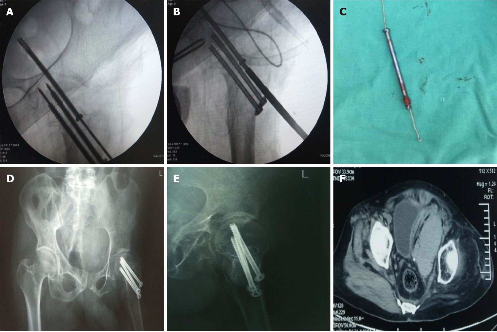
Figure 2 Intraoperative imaging and postoperative pelvic computed tomography.
A: Intraoperative fluoroscopy revealed that the cannulated screw was inserted too deeply; B: A self-made steel sternal wire was used to facilitate removal of the cannulated screw; C: Removal of the overlong cannulated screw and the self-made steel sternal wire with a hook; D: Postoperative posteroanterior radiograph; E: Postoperative lateral view; F: Postoperative pelvic computed tomography film showed effusion.
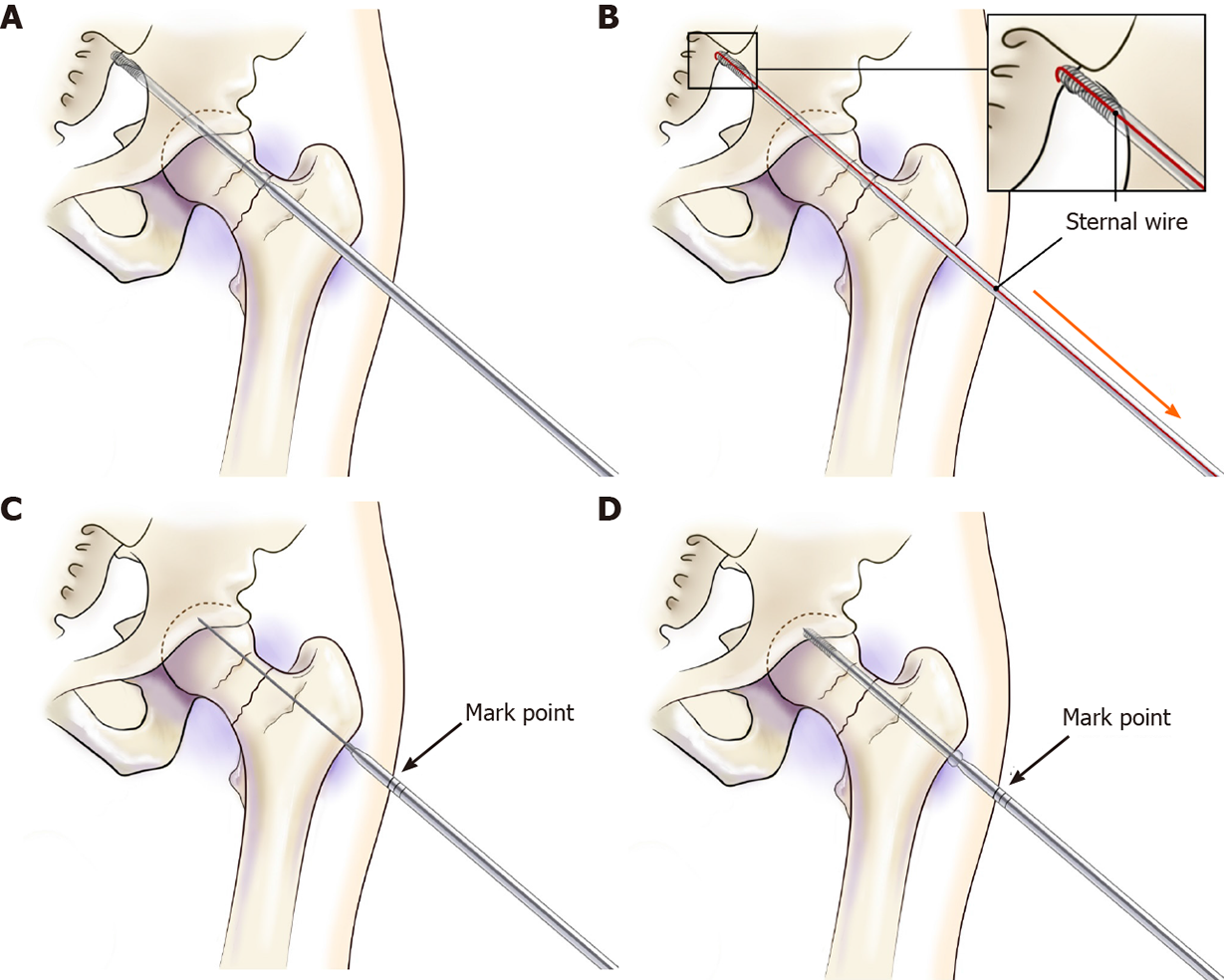
Figure 3 Process of removing the overly deep-placed cannulated screw and the use of the improved screwdriver for cannulated screws.
A: Inserting the excessively deep-placed screw; B: Using the self-made tool of steel sternal wire with bending and illustration for removal; C: Depth positioning using the scaled screwdriver before inserting the screw; D: Inserting the cannulated screw into the previously determined position with the scaled screwdriver.
- Citation: Yang ZH, Hou FS, Yin YS, Zhao L, Liang X. Minimally invasive removal of a deep-positioned cannulated screw from the femoral neck: A case report. World J Clin Cases 2021; 9(18): 4760-4764
- URL: https://www.wjgnet.com/2307-8960/full/v9/i18/4760.htm
- DOI: https://dx.doi.org/10.12998/wjcc.v9.i18.4760


