Copyright
©The Author(s) 2021.
World J Clin Cases. Jun 26, 2021; 9(18): 4728-4733
Published online Jun 26, 2021. doi: 10.12998/wjcc.v9.i18.4728
Published online Jun 26, 2021. doi: 10.12998/wjcc.v9.i18.4728
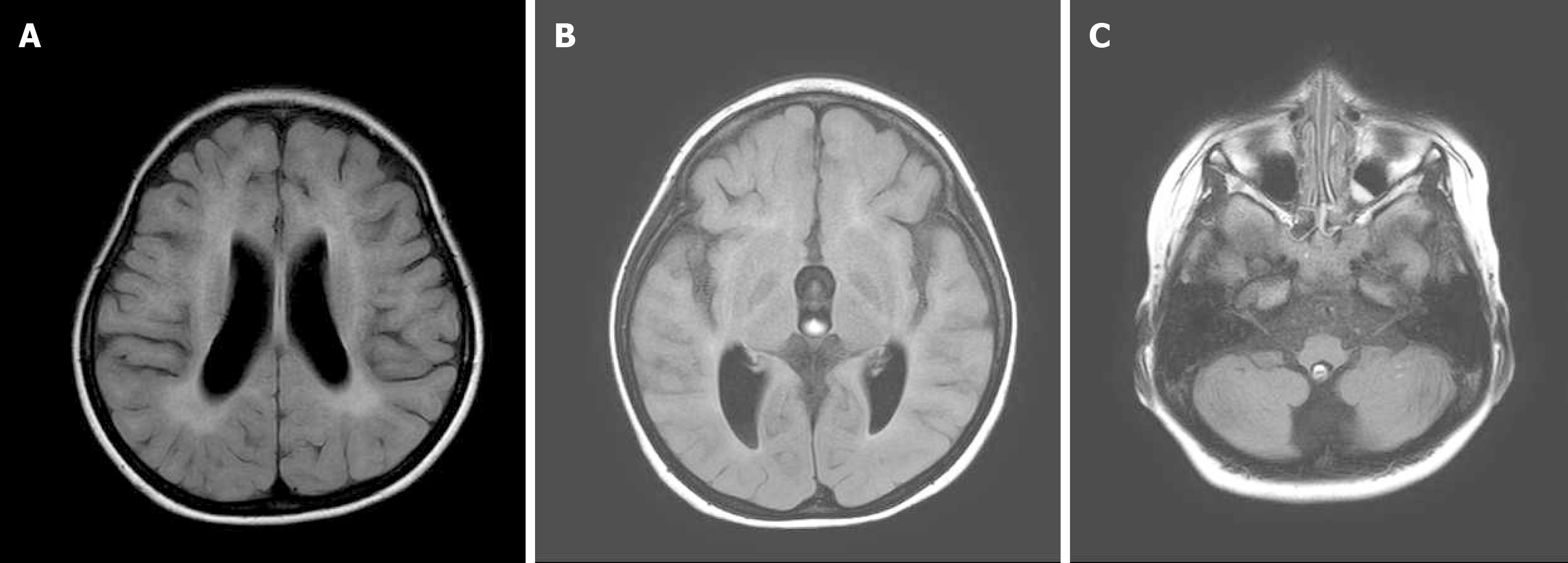
Figure 1 Magnetic resonance imaging of the brain without contrast medium.
A: Axial T2 FLAIR, leukodystrophy at the frontal subcortical and periventricular regions accompanied by a decreased amount of white matter; B and C: Axial T2 FLAIR, atrophic bilateral thalami, brain stem, and cerebellar hemispheres.
- Citation: Hsu LC, Chiang PY, Lin WP, Guo YH, Hsieh PC, Kuan TS, Lien WC, Lin YC. Botulinum toxin injection for Cockayne syndrome with muscle spasticity over bilateral lower limbs: A case report. World J Clin Cases 2021; 9(18): 4728-4733
- URL: https://www.wjgnet.com/2307-8960/full/v9/i18/4728.htm
- DOI: https://dx.doi.org/10.12998/wjcc.v9.i18.4728


