Copyright
©The Author(s) 2021.
World J Clin Cases. Jun 26, 2021; 9(18): 4607-4616
Published online Jun 26, 2021. doi: 10.12998/wjcc.v9.i18.4607
Published online Jun 26, 2021. doi: 10.12998/wjcc.v9.i18.4607
Figure 1 Measurement of spatial position parameters of the rib head/neck and transverse process space.
A: Adjustment of the observation angle: The spinous process line (dashed) is located in the middle of the bilateral pedicle lines (dotted), and the inferior border coincides with the anteroposterior border (solid line) of the superior thoracic vertebra; B: Parameter measurement: The vertical distance from the horizontal line of the inferior margin of the superior transverse process to the horizontal line of the superior margin of the inferior transverse process (dotted) is the width of the transverse process space; the vertical distance from the horizontal line of the highest point of the rib neck/head (dashed) to the horizontal line of the superior margin of the inferior transverse process (dotted) is the height of the rib neck/head protrusion. DP: The width of the intertransverse process space; DR: The height of the rib neck/head protrusion.
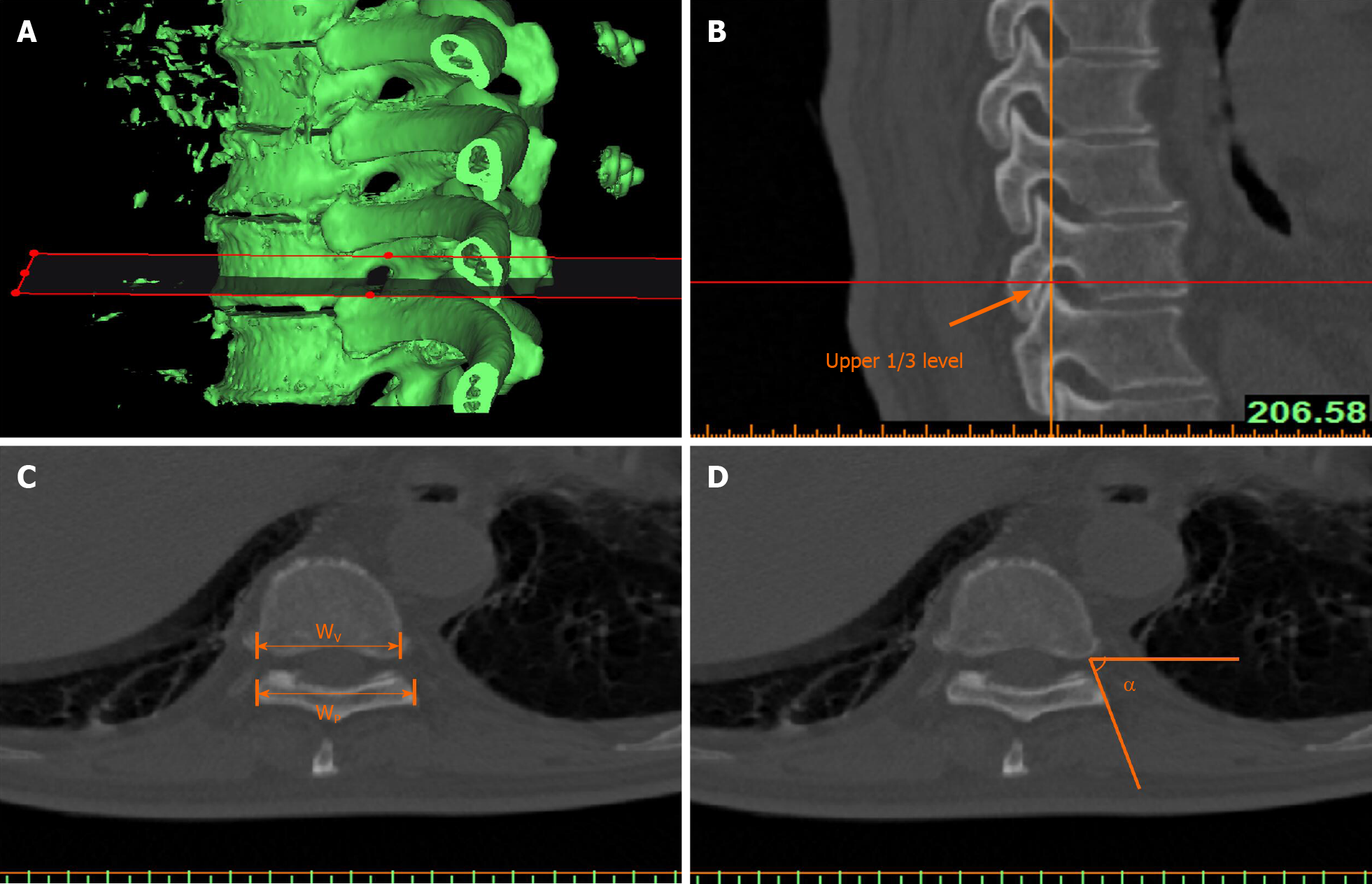
Figure 2 Measurement of parameters related to the intervertebral foramen.
A and B: The three-dimensional model of the thoracic vertebra was resliced parallel to the inferior border of the superior vertebral body of the intervertebral foramen, and the upper 1/3 level tomographic image of the intervertebral foramen was selected for parameter measurement; C and D: The width of the lateral border of the articular process/lamina; the width of the posterior border of the vertebral body; and the horizontal inclination angle from the lateral border of the articular process/lamina to the posterolateral border of the vertebral body. WP: The width of the lateral border of the articular process/lamina; WV: The width of the posterior border of the vertebral body; α: The horizontal inclination angle from the lateral border of the articular process/lamina to the posterolateral border of the vertebral body.
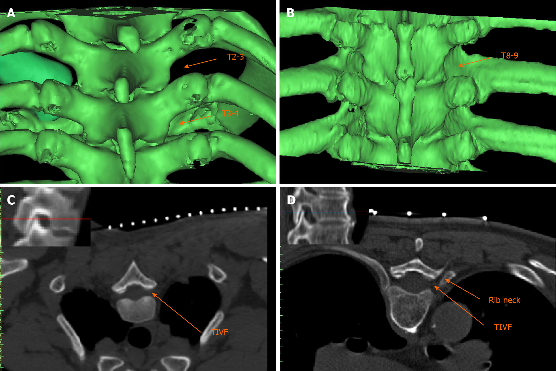
Figure 3 Spatial position of the rib head/neck and transverse process space.
A and B: The T2-3 intertransverse process space (ITPS) is not occluded by the rib neck/head, while the T3-4 ITPS is partially occluded, and the T8-9 ITPS is completely covered; C: The upper 1/3 level tomographic image of the T2-3 intervertebral foramen shows no occlusion by the corresponding rib neck/head; D: The upper 1/3 level tomographic image of the T8-9 intervertebral foramen shows occlusion by the corresponding rib neck; puncture of the intervertebral foramen would need to pass through the gap between the vertebral plate and the rib neck. TIVF: Thoracic intervertebral foramen.
- Citation: Wang R, Sun WW, Han Y, Fan XX, Pan XQ, Wang SC, Lu LJ. Observation and measurement of applied anatomical features for thoracic intervertebral foramen puncture on computed tomography images. World J Clin Cases 2021; 9(18): 4607-4616
- URL: https://www.wjgnet.com/2307-8960/full/v9/i18/4607.htm
- DOI: https://dx.doi.org/10.12998/wjcc.v9.i18.4607


