Copyright
©The Author(s) 2021.
World J Clin Cases. Jun 16, 2021; 9(17): 4423-4432
Published online Jun 16, 2021. doi: 10.12998/wjcc.v9.i17.4423
Published online Jun 16, 2021. doi: 10.12998/wjcc.v9.i17.4423
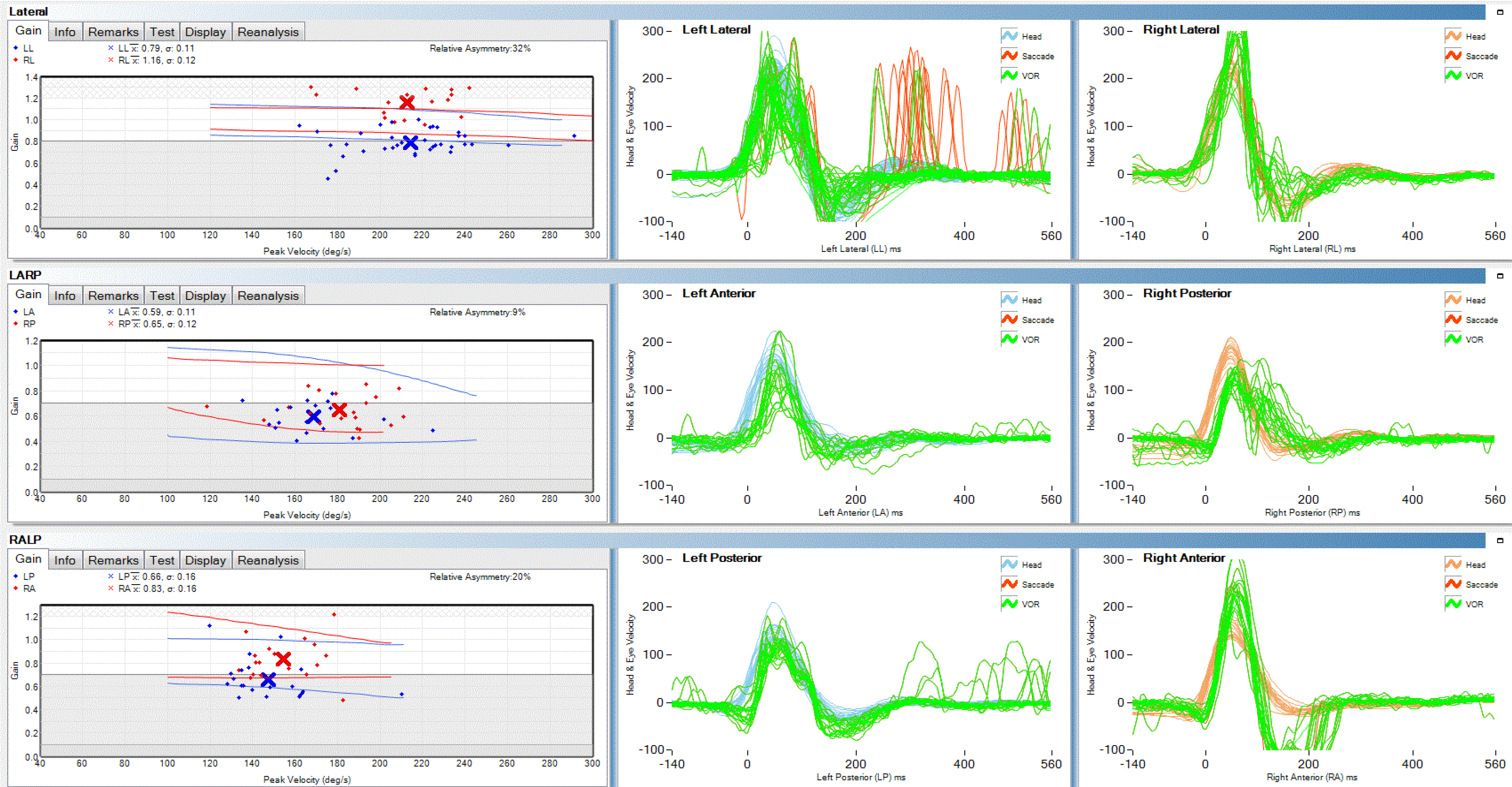
Figure 1 Video head pulse test suggested that the left semicircular canal gain (horizontal semicircular canal, posterior semicircular canal, and anterior semicircular canal) was slightly lower than the right gain, and the left horizontal semicircular canal was accompanied by glance.
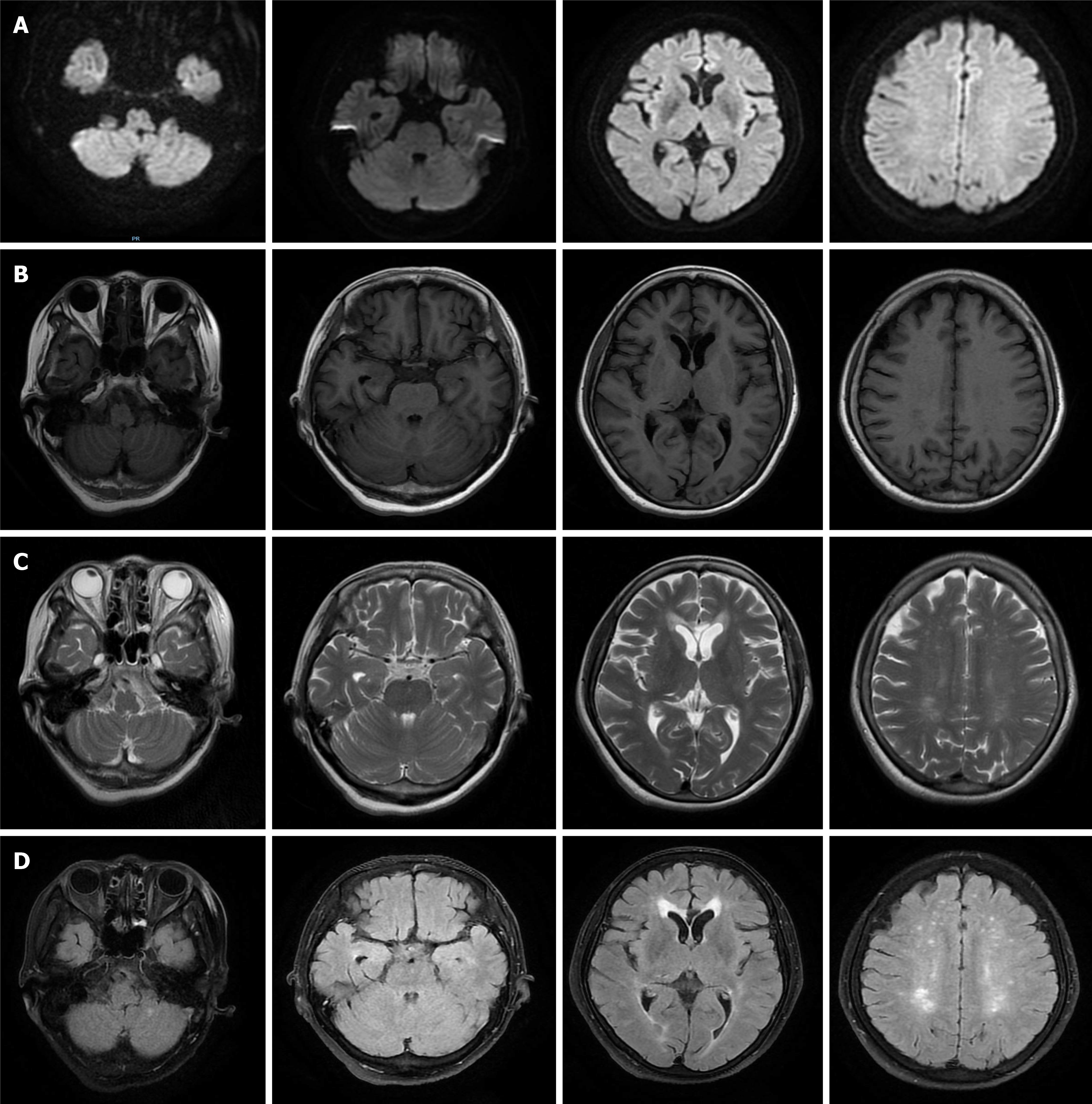
Figure 2 Magnetic resonance imaging showed multiple sub-frontal and parietal cortexes, semi-oval centers, and lateral ventricles with multiple spot-like and sheet-like lesions, equal signal on diffusion-weighted imaging, slightly longer signal on T1, longer signal on T2, and partially high signal on flair.
A: Diffusion-weighted imaging; B: T1; C: T2; D: Flair.
Figure 3 Contrast-enhanced computed tomography scan of the whole abdomen.
Multiple enlarged lymph nodes in the retroperitoneum and porta hepatis region and no obvious enhancement, which suggested a metastatic tumor. A: Retroperitoneum (orange arrow); B and C: Porta hepatis region (shown by orange arrows and orange circles).
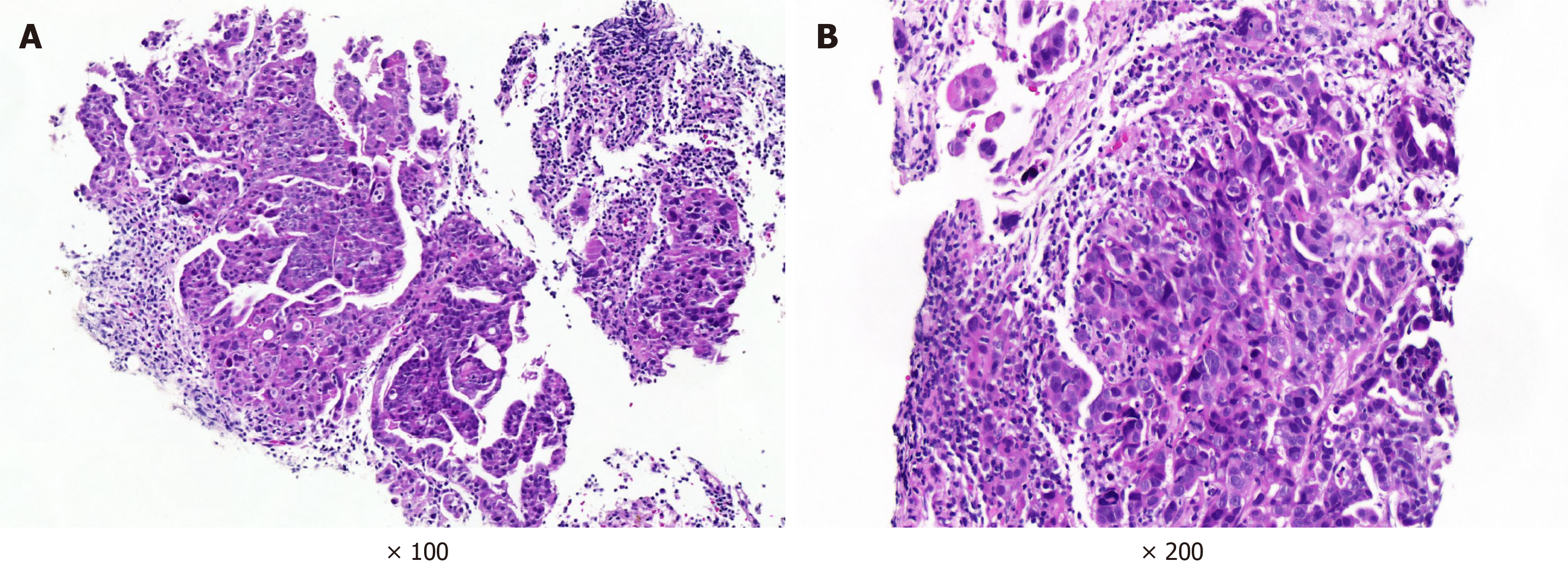
Figure 4 Lymph node pathology.
A: Left cervical lymph node pathology. Hematoxylin-eosin (HE) staining, × 100 magnification. The lymph nodes were infiltrated by atypical cells, and the atypical cells partly had an adenoid structure; B: Retroperitoneal lymph node pathology. HE staining, × 200 magnification. Atypical cells grew in nests, some of which had adenoid structures and infiltrating growth, with nuclear divisions visible
- Citation: Lou Y, Xu SH, Zhang SR, Shu QF, Liu XL. Anti-Yo antibody-positive paraneoplastic cerebellar degeneration in a patient with possible cholangiocarcinoma: A case report and review of the literature. World J Clin Cases 2021; 9(17): 4423-4432
- URL: https://www.wjgnet.com/2307-8960/full/v9/i17/4423.htm
- DOI: https://dx.doi.org/10.12998/wjcc.v9.i17.4423


