Copyright
©The Author(s) 2021.
World J Clin Cases. Jun 6, 2021; 9(16): 4046-4051
Published online Jun 6, 2021. doi: 10.12998/wjcc.v9.i16.4046
Published online Jun 6, 2021. doi: 10.12998/wjcc.v9.i16.4046
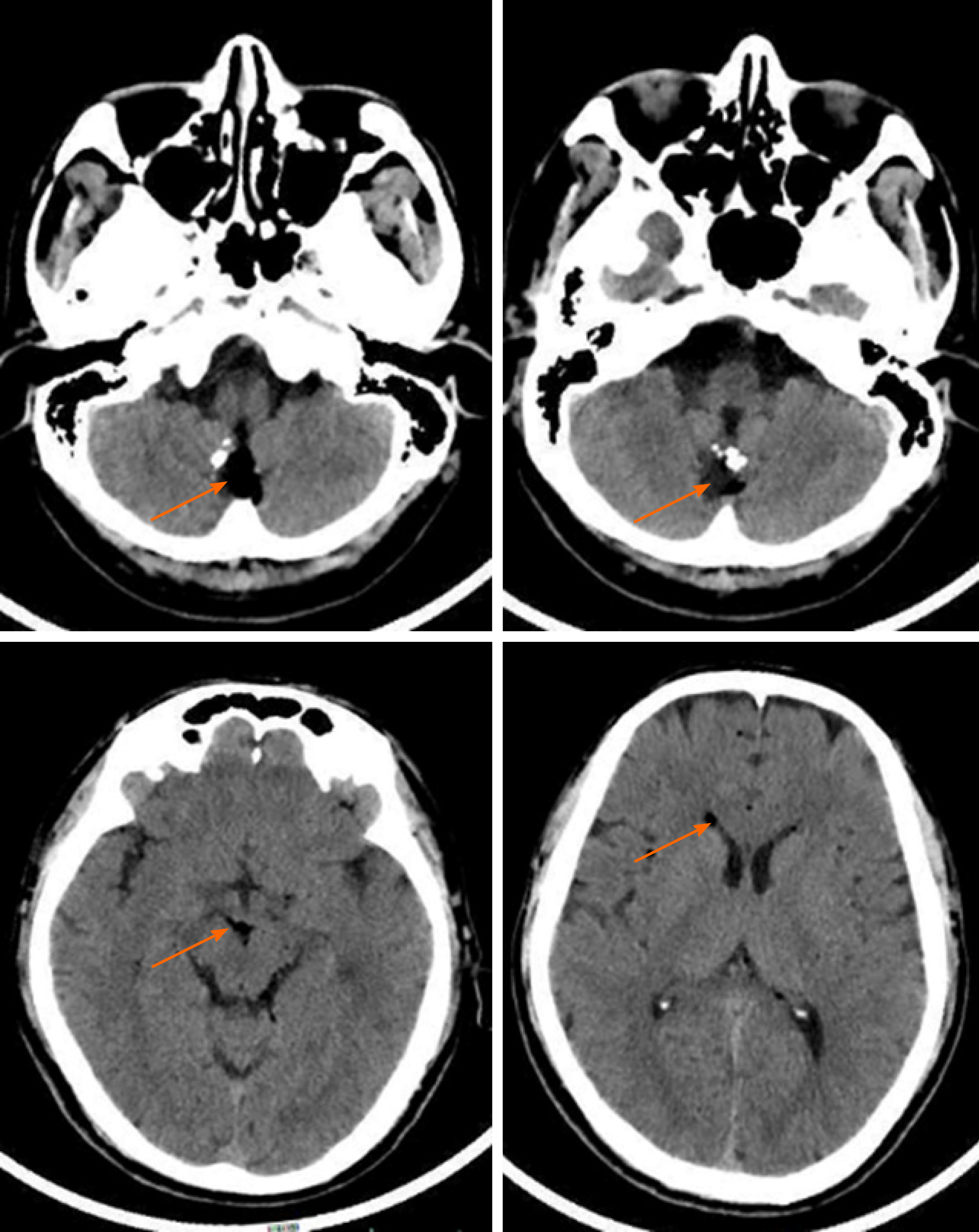
Figure 1 Cranial computed tomography scan showed a mixed-density lesion in the midline of the posterior fossa (arrow), with a fat-density area inside and calcified margin (arrow), as well as lipid droplet drifts in sulci, cisterns, and lateral ventricles (arrows).
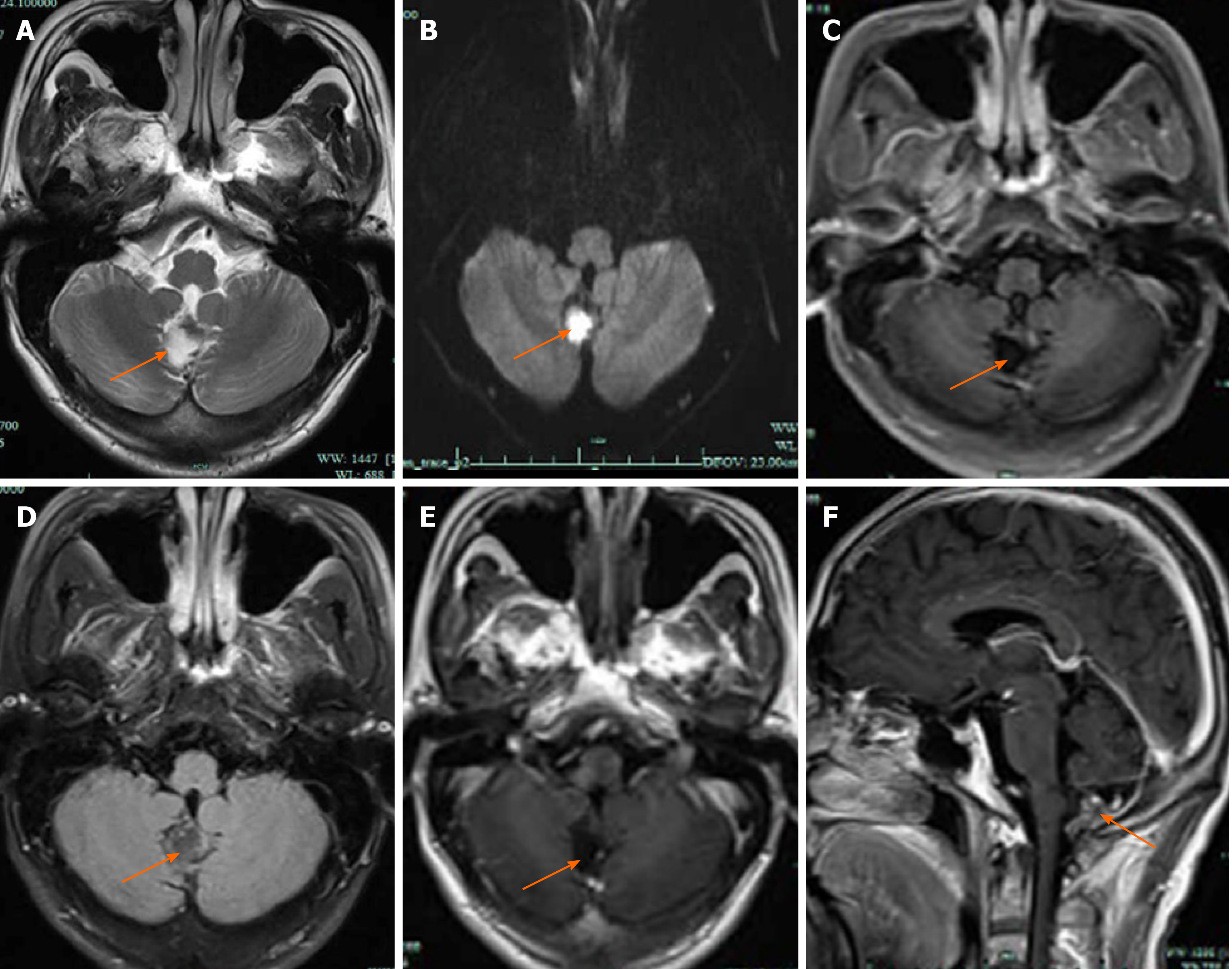
Figure 2 Cranial magnetic resonance imaging at 2 wk after injury.
A: The lesion presented hyperintensity on axial T2-weighted imaging (arrow); B: The lesion presented hyperintensity on diffusion weighted imaging (arrow); C: The lesion presented hypointensity on T1-fluid attenuated inversion recovery (FLAIR) (arrow); D: The lesion presented hypointensity on T2-FLAIR (arrow); E: No enhancement in the cystic lesion area was observed on axial T1-FLAIR (arrow); F: On the enhanced sagittal T1-FLAIR, no enhancement was observed in the cystic lesion, while the fat area presented hyperintensity and extended to the subarachnoid space (arrow).
- Citation: Zhang MH, Feng Q, Zhu HL, Lu H, Ding ZX, Feng B. Asymptomatic traumatic rupture of an intracranial dermoid cyst: A case report. World J Clin Cases 2021; 9(16): 4046-4051
- URL: https://www.wjgnet.com/2307-8960/full/v9/i16/4046.htm
- DOI: https://dx.doi.org/10.12998/wjcc.v9.i16.4046


